
As veterinarians, we are all too familiar with those devastating Friday afternoon emergencies. We are packing up for the weekend when a dog is rushed in with a hemoabdomen. The owner is devastated and says, “she was fine this morning.” A diagnostic workup confirms the patient has what looks like a ruptured splenic mass, with the most likely diagnosis being hemangiosarcoma.
Sadly, this is the way most cases of cancer in dogs are first detected–at a point when the disease is already advanced and the opportunity for cure is limited. These situations often leave families feeling blindsided and veterinarians feeling helpless. Something needs to change, and that change has arrived in the form of early cancer detection.
Background
Human medicine has shown early detection of cancer is the key to improved outcomes, which is why physicians recommend colonoscopies, mammograms, and prostate-specific antigen (PSA) tests to help detect cancer early. Recently, a new class of cancer screening tests have been adopted in humans that allow screening for dozens of types of cancer using a simple blood draw.1 These “multi-cancer early detection” or “MCED” tests use advanced genomic technology (called next-generation sequencing or NGS) to detect abnormal DNA in the blood (i.e. a “cancer signal”) that indicates cancer is present in the body.
The availability of “the blood test for cancer” or “liquid biopsy” represents one of the most promising new tools in the quest for early cancer detection. What is equally as exciting is that this game-changing technology is also now available for dogs.
In 2022, liquid biopsy testing using NGS was validated in an international, multi-center study of 1,100 dogs, the largest clinical validation study in veterinary oncology diagnostics.2 The test was proven to detect a cancer signal in blood for 30 different types of cancer and was particularly good at detecting cancers that are common and aggressive, such as lymphoma, hemangiosarcoma, and osteosarcoma. The false positive rate of the test was found to be only 1.5 percent, which is an important metric when considering implementing a screening test in large populations of dogs.
Additionally, test results are not impacted by medical conditions commonly encountered in routine practice, such as inflammation, infectious disease, autoimmune disease, and benign lumps and bumps.
Definition
Before we discuss how liquid biopsy can help with early cancer detection, it is important to define what is meant by “early detection.” When most people hear these words, they automatically think of cancer detection at an early disease stage (i.e. “early-stage detection”); however, early detection can also mean the identification of cancer before clinical signs develop (i.e. “preclinical detection” or “early clinical detection”).
It is well-established that dogs treated for cancer at an early disease stage demonstrate improved outcomes in most cancer types, including lymphoma,3,4 hemangiosarcoma,5 and osteosarcoma.6 among others.3,7–12 There is also a substantial literature base demonstrating improved outcomes for dogs treated for cancer prior to the onset of clinical signs, for instance in patients with: substage a lymphoma,13 non-ruptured hemangiosarcoma,5 absence of epistaxis for nasal tumors,14 and absence of cough/respiratory signs in lung tumors,15 among others.16–18
Impact on early detection
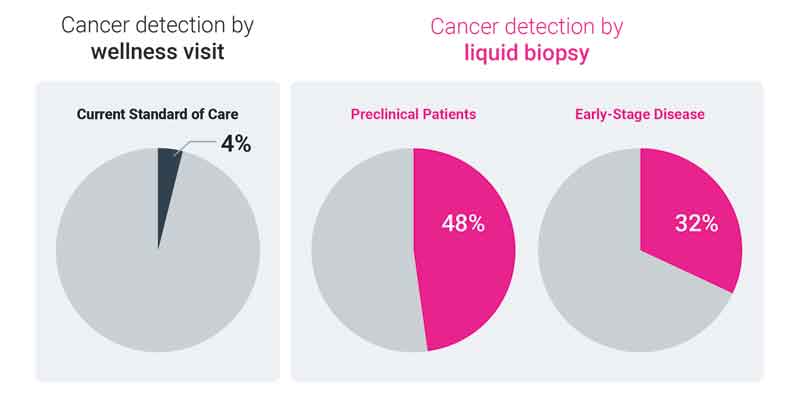
Currently, the standard of care for cancer detection in dogs is the annual or semi-annual wellness visit, in which the dog undergoes a thorough physical examination and routine bloodwork. This exam is critically important to monitoring a dog’s health and detecting a variety of conditions early (before the patient is sick), but what is the yield of this workup specifically for cancer detection?
In a recent study published in the Journal of Veterinary Internal Medicine (JVIM),19 medical records were reviewed for over 350 cancer-diagnosed dogs to determine how the disease first came to clinical attention. In this study, only four percent of dogs with cancer had their disease detected as the result of a routine wellness visit, prior to the development of clinical signs. On the other hand, liquid biopsy was able to detect a cancer signal in nearly half of preclinical patients (those that were asymptomatic) and in about one-third of patients with early-stage disease, demonstrating the potential for increased early cancer detection when liquid biopsy is added to a dog’s wellness visit (Figure 1).
Starting screening
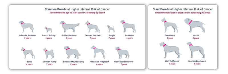
We now know that liquid biopsy can help identify cancer in dogs, so the next question becomes, “When should we start screening for cancer to catch the disease early?” To answer this, a recent study analyzed data from over 3,000 cancer-diagnosed dogs to determine the age at which they were diagnosed with the disease.20 The median age at cancer diagnosis in this population was approximately nine years; however, large dogs and dogs of certain breeds were found to be diagnosed with cancer younger, in some cases as early as age six. Taking into consideration decades of research (involving cancer latency periods, tumor doubling times, and cellular growth kinetics), the researchers arrived at a recommendation to start screening for cancer two years prior to the peak incidence of disease to increase the likelihood of early detection. So, as a general recommendation, all dogs should start cancer screening at age seven, but certain breeds may benefit from starting screening as early as age four (Figure 2).
Results
When implementing a new diagnostic tool in practice, particularly for a condition as impactful as cancer, important questions arise (i.e. “How often will I get a positive result?,” “How often is a positive result correct?,” “Will I be able to find the patient’s cancer?,” and “How long will the workup take?”). A study recently published in the Journal of the American Veterinary Medical Association (JAVMA) helps to answer these questions.21 In the study, 1,500 consecutive clinical samples submitted for liquid biopsy testing were analyzed. Approximately two-thirds of these real-world samples were submitted for cancer screening (in dogs with no current suspicion of cancer) and the majority of the remaining samples were submitted as an aid in diagnosis (for the workup of dogs with suspected cancer). A cancer signal (“positive result”) was detected in ~5 percent of cases submitted for screening, and in ~25 percent of cases submitted as an aid in diagnosis.
Patient outcomes were available for one-third of dogs in this study, and for patients that received a positive liquid biopsy result followed by a clinical workup, cancer was confirmed in ~90 percent of cases. These diagnoses were achieved quickly (a median of 11 days), typically using in-house capabilities; many of the cancer types detected in preclinical patients would not commonly be detected in an asymptomatic patient on wellness exam alone. The findings from this study provide further evidence that adding liquid biopsy to a dog’s wellness visit can help expand the number of cancer cases (and the range of cancer types) that can be detected preclinically.
Technology is helping us move the needle on early cancer detection in dogs. Detecting cancer early can have significant benefits for the patient, the owner, and the veterinary team. Early detection may mean more treatment options, improved quality (and in some cases, quantity) of life, and less financial burden to the owner.
In cases where fewer treatment options exist, it can give families the gift of time–time to pursue consultations on cancer care, time to decide what treatment plan fits best for their family, and most importantly, time to spend with their four-legged loved ones–whether that be completing a bucket list or preparing for proper end-of-life care for their beloved family member.
In all cases, it puts veterinarians and owners in control of the disease, rather than being controlled by a disease that can present in unexpected and sometimes life-threatening ways.
Andi Flory, DVM, DACVIM (Oncology), is a board-certified specialist in Medical Oncology. Dr. Flory graduated from The Ohio State University College of Veterinary Medicine and completed additional training at Florida Veterinary Specialists and Cornell University. Flory previously worked as an oncologist in the U.S. and Australia, until a little dog named Poppy changed the path of her career and led her to a passion for cancer genomics. After treating Poppy for advanced pancreatic cancer in 2019, Dr. Flory co-founded PetDx, to bring noninvasive cancer detection to veterinary medicine.
References
- Klein EA, Richards D, Cohn A, et al. Clinical validation of a targeted methylation-based multi-cancer early detection test using an independent validation set. Ann Oncol. 2021;32(9):1167-1177. doi:10.1016/j.annonc.2021.05.806 https://pubmed.ncbi.nlm.nih.gov/34176681/
- Flory A, Kruglyak KM, Tynan JA, et al. Clinical validation of a next-generation sequencing-based multi-cancer early detection “liquid biopsy” blood test in over 1,000 dogs using an independent testing set: The CANcer Detection in Dogs (CANDiD) study. Plos One. 2022;17(4):e0266623. doi:10.1371/journal.pone.0266623 https://journals.plos.org/plosone/article?id=10.1371/journal.pone.0266623
- Valli VE, Kass PH, Myint MS, Scott F. Canine Lymphomas. Vet Pathol. 2013;50(5):738-748. doi:10.1177/0300985813478210 https://pubmed.ncbi.nlm.nih.gov/23444036/
- Lautscham EM, Kessler M, Ernst T, Willimzig L, Neiger R. Comparison of a CHOP-LAsp-based protocol with and without maintenance for canine multicentric lymphoma. Vet Rec. 2017;180(12):303. doi:10.1136/vr.104077 https://pubmed.ncbi.nlm.nih.gov/28100766/
- Treggiari E, Borrego JF, Gramer I, et al. Retrospective comparison of first‐line adjuvant anthracycline vs metronomic‐based chemotherapy protocols in the treatment of stage I and II canine splenic haemangiosarcoma. Vet Comp Oncol. 2020;18(1):43-51. doi:10.1111/vco.12548 https://pubmed.ncbi.nlm.nih.gov/31648405/
- Spodnick GJ, Berg J, Rand WM, et al. Prognosis for dogs with appendicular osteosarcoma treated by amputation alone: 162 cases (1978-1988). J Am Vet Med Assoc. 1992;200(7):995-999. https://pubmed.ncbi.nlm.nih.gov/1577656/
- Horta RS, Lavalle GE, Monteiro LN, Souza MCC, Cassali GD, Araújo RB. Assessment of Canine Mast Cell Tumor Mortality Risk Based on Clinical, Histologic, Immunohistochemical, and Molecular Features. Vet Pathol. 2018;55(2):212-223. doi:10.1177/0300985817747325 https://pubmed.ncbi.nlm.nih.gov/29338615/
- Linden D, Liptak JM, Vinayak A, et al. Outcomes and prognostic variables associated with primary abdominal visceral soft tissue sarcomas in dogs: A Veterinary Society of Surgical Oncology retrospective study. Vet Comp Oncol. 2019;17(3):265-270. doi:10.1111/vco.12456 https://pubmed.ncbi.nlm.nih.gov/30666781/
- Tierce R, Martin T, Hughes KL, et al. Response of Canine Soft Tissue Sarcoma to Stereotactic Body Radiotherapy. Radiat Res. 2021;196(6):587-601. doi:10.1667/rade-20-00271.1 https://pubmed.ncbi.nlm.nih.gov/34473832/
- Turek M, LaDue T, Looper J, et al. Multimodality treatment including ONCEPT for canine oral melanoma: A retrospective analysis of 131 dogs. Vet Radiol Ultrasoun. Published online 2020. doi:10.1111/vru.12860 https://onlinelibrary.wiley.com/doi/abs/10.1111/vru.12860
- Sorenmo KU, Rasotto R, Zappulli V, Goldschmidt MH. Development, Anatomy, Histology, Lymphatic Drainage, Clinical Features, and Cell Differentiation Markers of Canine Mammary Gland Neoplasms. Vet Pathol. 2011;48(1):85-97. doi:10.1177/0300985810389480 https://journals.sagepub.com/doi/10.1177/0300985810389480
- Polton GA, Brearley MJ. Clinical Stage, Therapy, and Prognosis in Canine Anal Sac Gland Carcinoma. J Vet Intern Med. 2007;21(2):274. doi:10.1892/0891-6640(2007)21[274:cstapi]2.0.co;2 https://pubmed.ncbi.nlm.nih.gov/17427388/
- Jagielski D, Lechowski R, Hoffmann-Jagielska M, Winiarczyk S. A Retrospective Study of the Incidence and Prognostic Factors of Multicentric Lymphoma in Dogs (1998–2000). J Vet Medicine Ser. 2002;49(8):419-424. doi:10.1046/j.1439-0442.2002.00458.x https://pubmed.ncbi.nlm.nih.gov/12450190/
- Rassnick KM, Goldkamp CE, Erb HN, et al. Evaluation of factors associated with survival in dogs with untreated nasal carcinomas: 139 cases (1993–2003). J Am Vet Med Assoc. 2006;229(3):401-406. doi:10.2460/javma.229.3.401 https://avmajournals.avma.org/view/journals/javma/229/3/javma.229.3.401.xml
- McNiel EA, Ogilvie GK, Powers BE, Hutchinson JM, Salman MD, Withrow SJ. Evaluation of prognostic factors for dogs with primary lung tumors: 67 cases (1985-1992). J Am Vet Med Assoc. 1997;11(211):1422-1427. https://pubmed.ncbi.nlm.nih.gov/9394893/
- Debreuque M, Fornel PD, David I, et al. Definitive-intent uniform megavoltage fractioned radiotherapy protocol for presumed canine intracranial gliomas: retrospective analysis of survival and prognostic factors in 38 cases (2013–2019). Bmc Vet Res. 2020;16(1):412. doi:10.1186/s12917-020-02614-x https://pubmed.ncbi.nlm.nih.gov/33129320/
- Pecceu E, Varela JCS, Handel I, Piccinelli C, Milne E, Lawrence J. Ultrasound is a poor predictor of early or overt liver or spleen metastasis in dogs with high‐risk mast cell tumours. Vet Comp Oncol. Published online 2020. doi:10.1111/vco.12563 https://pubmed.ncbi.nlm.nih.gov/31863546/
- Treggiari E, Pellin MA, Romanelli G, et al. Tonsillar carcinoma in dogs: Treatment outcome and potential prognostic factors in 123 cases. J Vet Intern Med. Published online 2023. doi:10.1111/jvim.16623 https://onlinelibrary.wiley.com/doi/full/10.1111/jvim.16623
- Flory A, McLennan L, Peet B, et al. Cancer detection in clinical practice and using blood‐based liquid biopsy: A retrospective audit of over 350 dogs. J Vet Intern Med. 2023;37(1):258-267. doi:10.1111/jvim.16616 https://pubmed.ncbi.nlm.nih.gov/36661398/
- Rafalko JM, Kruglyak KM, McCleary-Wheeler AL, et al. Age at cancer diagnosis by breed, weight, sex, and cancer type in a cohort of more than 3,000 dogs: Determining the optimal age to initiate cancer screening in canine patients. Plos One. 2023;18(2):e0280795. doi:10.1371/journal.pone.0280795 https://journals.plos.org/plosone/article?id=10.1371/journal.pone.0280795
- O’Kell A, Lytle K, Cohen T, et al. Clinical experience with next-generation sequencing-based liquid biopsy testing for cancer detection in dogs: A review of 1,500 consecutive clinical cases. J Am Vet Med Assoc. Published online 2023. https://doi.org/10.2460/javma.22.11.0526