When looking at building or updating a veterinary dental suite, there will be big decisions on fairly major investments for the practice. Similar to other large purchases, such as a car, you need to start out with an idea of what features are necessary, which extras to consider, and the budget to work with. Many equipment manufacturers and distributors have a wide range of options available, so working with a trusted provider or even bargain shopping at a major conference may be helpful.
Dental tables
While it is certainly possible to work on a wet table with a cabinet type base (preferably with some leg room at the end of the table [Figure 1]) being able to adjust the height of the table is better for ergonomic considerations. This will help with team members of different heights and allow standing for some of the procedures or sitting for others.
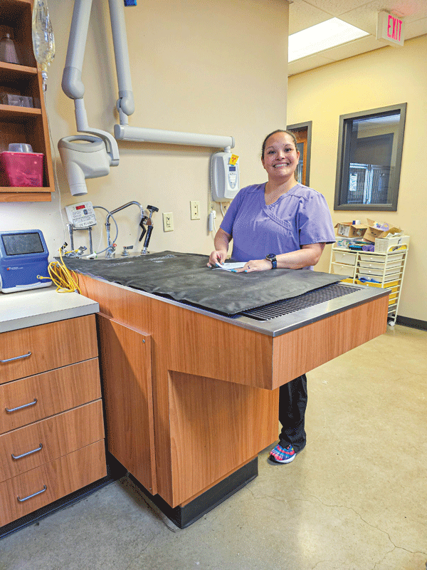
but no adjustability on height. It is important to have space to provide room for
a seated operator. Photo courtesy Dr. Lobprise
A table that can be lowered close to the floor may also be useful for larger dogs. Anti-fatigue floor mats should be used to provide further support and comfort to team members who stand for long periods of time. These should be easy to move and clean when not used or as necessary.
Since dental procedures require a water source, there must be a method for managing or collecting the water waste. It is optimal to keep the water away from the patient as much as possible to avoid hypothermia. There are many styles of wet tables with a tub underneath and a rack or grate on top to allow fluid drainage. If directly connected to a water source and drainage, cleaning up can be done easily. If not connected, often a collection device or bucket under the table will catch the fluid, so monitoring the amount of fluid and emptying the device is needed.
Chairs
As with the tables, appropriate, adjustable chairs that encourage a more natural and comfortable posture for the operators can help avoid musculoskeletal disorders. The padded seat may be flat, contoured or saddle-shaped depending on operator preference (Figure 2), with the ability to place the feet on the floor with a slight bend in the knee. If a back rest is used, it should be adjusted to contact the curve of the lower back. Seat tilt adjustment is optimal in the 10- to 15-degree range, and some chairs allow a “sit-to-stand” configuration.1
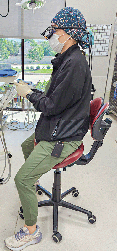
better ergonomic posture and has a sit-to-stand
feature to allow for easy changes in position. Photo courtesy Shannon VanTrease
Lighting and visualization
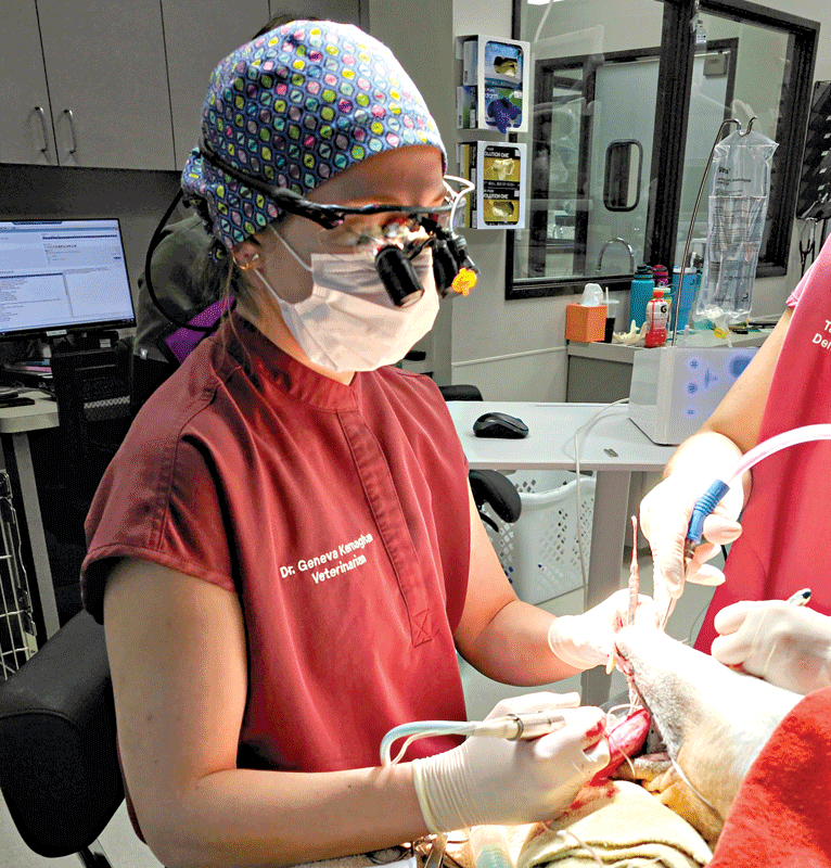
Good visualization is critical for dental procedures, and overhead lights should be easily adjustable for both position and light intensity. Optimally, a light source that moves with the operator will provide continuous illumination in the field of operation, and these are often paired with magnification loupes. Not only do loupes help with magnification and light, but they can allow a more ergonomically supportive posture.1
As with other practice equipment, ensure you take loupes for a trial run before purchase to find the right fit for you. Traditional through-the-lens loupes are most frequently used, with slight forward neck inflection of 10-20 degrees that is reasonable for most operators (Figure 3).
Operator support
Accompanying appropriate dental tables, chairs and loupes, operator protection is critical. Eye covering, masks, and gloves help protect the technician team and veterinarian from aerosolized bacteria and debris. Adequate ventilation for the area should be provided beyond anesthetic scavenging systems.
Dental delivery unit
Determining the basic must-haves, the options, and your budget are important starting points for selecting a dental delivery unit. The delivery of compressed air is typically from an in-unit compressor, or remote compressor if multiple units are used. Compressed nitrogen may also be used in some cases.
- Compressor selection and maintenance are key to selecting the right unit for your practice and keeping it working optimally for years to come. Some are oil-cooled and require regular maintenance, and some are air-cooled, requiring filters and cleaning.
The compressed air is used to drive the handpieces used on the unit. At minimum, a high-speed handpiece, a low-speed handpiece, and an air/water syringe are recommended. A vacuum attachment can be a very helpful option (Figure 4). The high-speed handpiece should always have a bur or blank inserted and run at the full speed of 300,000 to 400,000 RPM with adequate water spray. In most units, this is achieved with approximately 40 PSI delivered to the handpiece. Both the water spray and PSI can be adjusted. These handpieces need regular (at least daily) cleaning, sterilization, and lubrication2 (Figure 5).
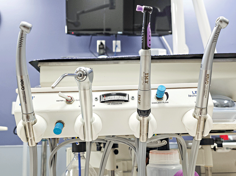
Figure 4. High-speed unit (remote compressor for air source) with multiple handpieces (from left): high-speed handpiece; air-water syringe; low-speed handpiece; second high-speed handpiece, which connection can also be used for a vacuum handpiece. Photo courtesy Shannon VanTrease The low-speed handpiece on the unit can run up to 20,000 RPM, but with polishing (the most common use), it should be at around 3000 RPM, similar to the speeds used by micro-motor polishing units. There is no water spray with this, so care must be used not to generate excessive heat with higher speeds or operator pressure. Cleaning and maintenance are also required.
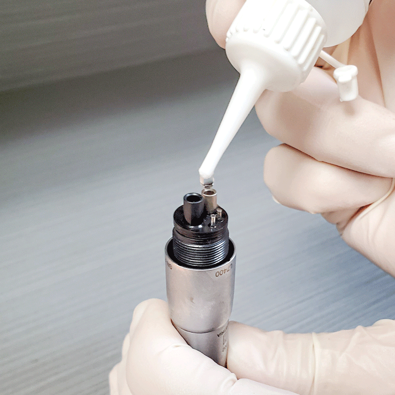
Figure 5. High-speed handpieces need daily cleaning and lubrication; the manufacturer provided oil should be placed in the larger of the two side-by-side holes each day. Photo courtesy Shannon VanTrease - As part of the dental unit, or as a separate piece, an ultrasonic scaler does not rely on compressed air to function. Electrical current stimulates the scaler to convert sound waves into mechanical vibration that removes debris off the teeth. The distilled water flow not only keeps the scaler tip cooled, but it also creates cavitation bubbles that help disrupt bacterial cell membranes.
Magnetostrictive scalers have metal rods or sheets in stacks that vibrate in an elliptical fashion; piezoelectric crystals are activated in a linear fashion. With either type, the side (and not the tip) of the scaler should be used on the tooth surface. Water spray must be able to reach the tip as a coolant, and that can be the guideline as to whether a tip can be used subgingivally or not.
In addition to regular cleaning, the scaler tips should be measured after each use; when wear is below the 2-3 mm mark on the guide provided by the manufacturer, it should be replaced. Dropping the handpieces can also separate the magnetostrictive stacks or shatter the piezoelectric crystals.
Radiograph unit
The dental radiographic generator has several options, including wall-mounted, movable-stands, and hand-held units. Radiographic variables of kVp (kilovolt peak) and mA (milliamperage) typically do not need adjustment, and minimal variations of the time of exposure can even be adjusted by the unit, based on patient size, type of film/sensor and tooth/teeth to be imaged.3 Selecting a radiograph unit may be dependent on the area in which you work and how you need images to be captured.
Digital dental radiography is commonly used in practices, often with a digital sensor that provides an immediate image on the computer screen (direct digital radiography). While this is faster, the thicker size 2 sensor is sometimes challenging to position in small patients, and replacement of a damaged sensor can be expensive.
Semi-direct digital radiography or computed radiography utilizes photo-stimulable phosphor plate that provide additional sizes of plates that are much thinner than digital radiography sensors, flexible, and less expensive. It does take several seconds to be scanned, so the image can be generated and seen on the computer screen. A good practice hack is getting an inexpensive TV screen, connecting it to the computer, and mounting it on the wall to view the enlarged images easily (Figure 6).
Patient support and care
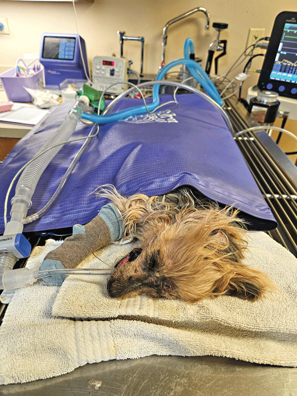
Appropriate dental procedural care requires using general anesthesia to perform complete dental examinations, take dental radiographs, and perform procedures. Even with total intravenous anesthesia, cuffed endotracheal tubes are essential to protect the patient’s airway from aerosolized particles and debris and to provide oxygen support.
Since water delivery is essential, our patients often get their heads wet, which can decrease their body temperature. Thermal regulation is even more challenging in smaller and older patients, so providing devices that help maintain body temperature is critical in a dental practice.
Passive warming (blankets, towels, socks for the feet) are often supplemented with active warming devices, such as forced warm air blankets, water-circulating blankets, and air-free conductive warming (Figure 7).
Active surface warming with heating pads or lamps should be used very carefully, especially in cats. Warming IV fluids is also an option.
Since general anesthesia is needed to perform complete dental examinations, patient monitoring is also critical. While the best monitor is a good technician (able to assess many of the physical parameters manually), systems are available to assess temperature, respiration rate, heart rate, EKG, blood pressure, pulse oximeter, and end-tidal CO2. Do not forget to continue monitoring during the recovery phase with regular visual assessment. This can now be enhanced by using a vital signs remote monitor (Doppler radar technology) to detect temperature, respiration and heart rate with live video and accessed with any smart device.
Summary
Providing a good foundation with appropriate operatory equipment and units can enhance your team’s ability to provide optimal dental care for your patients. In the succeeding parts of this feature, authors will tackle dental instruments (part two) and dental materials and supplies (part three).
Heidi Lobprise, DVM, DAVDC, is a 1983 Texas A&M University graduate. She became board-certified in dentistry in 1993. After 10 years in the industry, Dr. Lobprise returned to dental specialty practice in 2014 and has since “semi-retired” in Kerrville, Texas. Lobprise is the author/co-author of three dental texts, writing many chapters and articles.
Shannon VanTrease, a dental practice manager (FMVESC), has been in the veterinary field for more than 20 years and has a wide variety of experience, including emergency medicine, companion animal practice, large animal medicine, with more than 13 years in veterinary dentistry. VanTrease’s interest in veterinary dentistry began during his time working for Robert Wiggs, DVM, DAVDC, and then Heidi Lobprise, DVM, DAVDC.
References
- Ergonomics and Posture Guidelines for Oral Health Professionals. FDI_HSDW_ergonomics_and_posture_guidelines_eng_2021.pdf (fdiworlddental.org). Accessed 04-01-24.
- Holmstrom SE. Dental instruments and equipment. Veterinary Dentistry: A Team Approach. 3rd ed. Elsevier; 2018:72-110.
- Niemiec B. Oral Radiology and Imaging. In: In: Lobprise HL, Dodd JR, eds. Wiggs’ Veterinary Dentistry: Principles and Practice. Wiley Blackwell; 2019: 41-62.