Photos courtesy John R. Lewis
Approaching is the 19th anniversary of one of the most interesting cases I have ever seen.
I had just finished my residency in dentistry and oral surgery in July 2004 at the Ryan Veterinary Hospital at the University of Pennsylvania (VHUP). Thereafter, I stayed on as a lecturer, which proved to be a supportive environment as I studied for my board examination in spring 2005.
The doctors of the Dentistry and Oral Surgery Service at VHUP took turns being on call during weekends so the doctors in emergency service had someone available if they had questions about patients that presented with oral and maxillofacial conditions through the emergency service.
Though there are not a large amount of true dental emergencies, occasionally we would be called in for a procedure that ideally shouldn’t wait until Monday. Many weekend procedures included cases such as jaw fractures, dislocated TMJs, traumatically avulsed teeth that owners hoped to save, etc. Here’s a blast from the past. Do you remember Nextel phones? That was the communication method among clinicians, nurses, and administration at VHUP in the early 2000s. For readers who may not be old enough to recall the Nextel era, the phone functioned as a cellular phone, but it also had a side button that allowed it to be used as a walkie-talkie.
I was on call one Saturday in mid-November 2004 when I received a Nextel from Dr. Dan Hume in the Emergency Service at Penn.
Dr. Hume: “Dr. Lewis, are you available?”
Dr. Lewis: “Yes, go ahead.”
Dr. Hume: “We’ve got an approximately two-year-old mixed breed dog here that presented with trauma to the bridge of her nose. The dog, Alice, was enjoying herself on a farm when Charlotte, her owner, noticed that Alice had wandered away, and went looking for her. She found Alice hiding just outside the house with a fresh wound at the bridge of the nose. She was assessed at her primary care veterinarian, who noted she was mentally appropriate with no other apparent trauma. Silver sulfadiazine was placed on the wound, and she was sent to the ER at VHUP.”
Dr. Lewis: “OK, Dan, I’ll be there in about half an hour.”
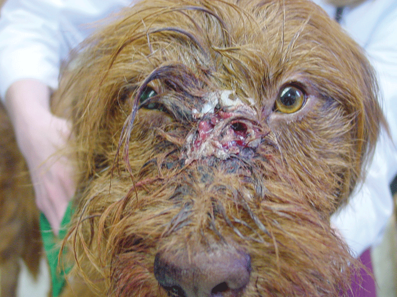
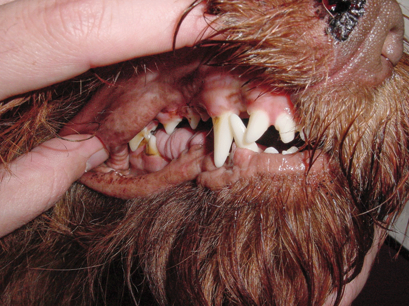
When I arrived in the ER, Alice was bright, alert, wagging her tail, and encouraging every passerby to stop and greet her. The most obvious abnormality was the wound at the bridge of her nose, which was not actively bleeding. However, dried blood was noted at the right nostril, and Alice’s right pupil was mydriatic, compared to the normal looking left pupil (Figure 1). I pulled her upper lips out of the way to check the occlusion. Occlusion was almost completely normal, except for a slight shift of the lower jaw to the right (Figure 2).
Alice was too painful to try to open her mouth while she was awake, but I could see there was significant swelling and bruising in the caudal right oropharynx.
We decided to place Alice under anesthesia to perform an oral exam and obtain imaging. During intubation, the anesthesia technician reported there was resistance upon opening the mouth and a decreased range of motion. Severe right pharyngeal swelling and bruising was confirmed. Resistance was significant when attempting to open the mouth, so we avoided prying the mouth open until we could obtain imaging, which turned out to be a good decision.
We started with radiographs of Alice’s head and neck. The radiology department at VHUP was located on the first floor, and my office and the operatory were on the third floor. While the radiographs were being obtained,
I ran upstairs to call Charlotte with an update on Alice. Just at that moment, Irene from the radiology department called via Nextel.
Irene: “Dr. Lewis are you available?”
Me: “Yes, go ahead.”
Irene: “You need to come to radiology to see these radiographs!”
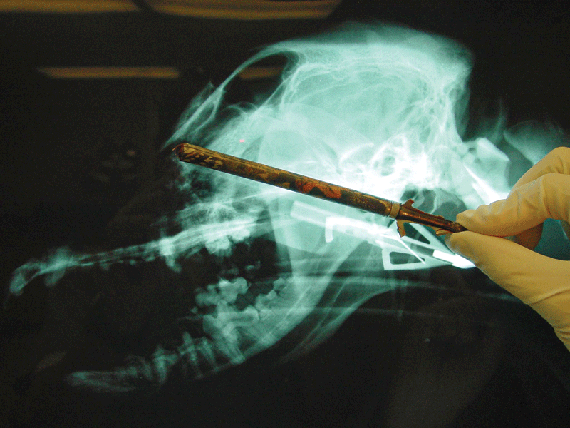
Remember this case occurred 19 years ago, at a time before we had digital radiography, so hard copy films were posted on the view box. My curiosity was piqued, I rushed down two flights of steps to radiology. I walked into the viewing room, and there it was: the reason was obvious for Alice’s wound and pharyngeal swelling. The wound was created by a hunting arrow, and approximately six inches of its shaft traveled from the entry point at the bridge of the nose and lodged just medial to the right temporomandibular joint (TMJ), which had a condylar fracture (Figure 3). The remaining portion of the arrow must have broken off at the level of the skin. It was the type of arrow that had three razor sharp prongs at the tip, one of which broke off on impact and was lodged in the TMJ area.
How was this situation dealt with? Tune in next month for lessons learned of the team effort that allowed Alice to live a long, productive life.
John Lewis, VMD, DAVDC, FF-OMFS, practices and teaches at Veterinary Dentistry Specialists and Silo Academy Education Center, both located in Chadds Ford, Pa.