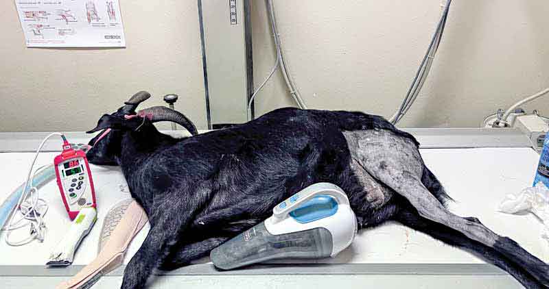
Photos courtesy Jeff Mayo
The most common pathogen involved in small animal surgical site infections today is the methicillin-resistant Staphylococcus pseudintermedius (MRSP). The overall carrier rate of MRSP is approximately 4.4 percent, with a 14 times higher infection rate in part due to its ability to develop a biofilm. This, and other pathogens, are found in the normal commensal organisms of the canine skin appendages. Typical skin infection rates for most elective surgical procedures run approximately three percent to five percent, though are much higher in TPLO surgeries, reported up to 21-28 percent, becoming a more difficult problem to combat.
Surgical site infections
Surgical site infections (SSIs) are defined by the Centers for Disease Control and Prevention (CDC) as: superficial incisional (less than 30 days), deep incisional (30 days to 12 months), or organ space (30 days to 12 months).
Surgical wounds are classified as clean, clean-contaminated, contaminated and dirty, and preventable or non-preventable. Acute SSIs may arise from other comorbid infections prior to the procedure, such as urinary tract infections (UTIs) or pyoderma. Chronic infections may result from the hematogenous spread from other distant areas such as dental disease or endocarditis. There is concern reported infection rates are not accurate as many may be treated by doctors not involved in the initial treatment of the patient, or the incident may not be reported in the record accurately.
Many causes of increased susceptibility to SSIs are cited in the literature.1-3 An SaO2 < 90 percent, or a blood pressure less than 60, increase the risk of SSI by 27 times. Use of propofol as an induction agent increases the susceptibility to infection by 38 percent. Prolonged anesthetic times, increased number of people in the operating room, and clipping the hair longer than four hours before surgery may also make patients more susceptible to SSIs. In TPLO surgery, use of non-locking plates may lead to increased instability of the osteotomy site and necrosis around the screw-holes, leading to potential SSI, though a more recent study refutes this idea.4 Using skin staples increases the chances of infection from approximately three percent to five percent, while the use of triclosan-impregnated sutures may decrease SSIs.
Hospital-acquired infections (HAIs) occur most commonly during the placement of medical devices, such as bone plates, intramedullary pins, IV, or urinary catheters. Long procedure times, or underdeveloped skill may contribute to increased infection rates. Clothing worn by hospital staff cultured positive for MRSP 14 percent, and methicillin-resistant Staphylococcus aureus (MRSA) at a rate of 3.5 percent.5 Commonly identified sources of infection included the patient gurney, the radiology table (most common), and the staffs’ hands that handled the patient after surgery.
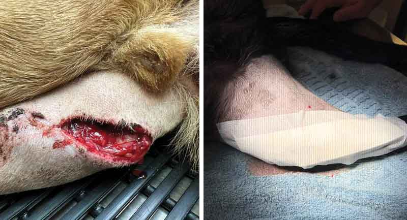
Right: Immediate post-operative wound bandaging minimizes surgical infections.
Deterrence methods
Deterrence methods of SSIs are cited in the literature.6,7 Using triclosan-impregnated suture and intradermal closures might be a better choice than skin staples. Iodine-impregnated sheets applied to the patient during surgery and use of impervious barriers decrease chances of SSIs. Double-gloving or using orthopedic surgery gloves minimize the chances of glove puncture during a procedure, although gloves should still be changed after draping the patient, and after 60 minutes of surgery. Application of an antimicrobial cream and a light bandage before leaving the operating table is a suggested practice to minimize wound contamination while still in the hospital.
- Patient selection. Proper patient selection is important to minimize the chance of an SSI. Older patients in poor health, or obese intact males, incurred higher SSIs in one study8 Patients with concurrent comorbidities may likely increase the odds of acquiring an SSI. The use of orthopedic implants dramatically increases the opportunity for the formation of an SSI and a biofilm, making infection resolution difficult. Knowing the potential epidemiology of the offending bacteria prior to surgery and pre-planning antimicrobial prophylaxis plan is becoming increasingly difficult as resistant rates increase to current antibiotics in use.
- Antimicrobial prophylaxis (AMP) The CDC currently recommends AMP in human medicine be discontinued 24 hours after surgery.9 AMP continues to be a great area of debate in veterinary medicine as there are currently no standardized guidelines. Most proposed protocols include using various dosages of cephalexin, with most SSIs involving organisms that are resistant to that drug. Other methods of prevention, such as frequent hand washing, wearing gloves when handling patients, and keeping the wounds covered at least until discharge from the hospital should preclude inappropriate use of AMP. The literature does seem to agree that some sort of AMP is appropriate when performing orthopedic procedures involving bone plate implants in dogs.10-12
- Surgeon preparation. This step should begin with appropriate clothing, surgical scrubs, hat, mask, sterile gown, and a three-minute scrub of the hands with an appropriate purpose-based and dedicated hand scrub, preferably in one that is foot-activated to minimize contamination. Hand rubs—chlorhexidine and alcohol-based—are becoming more popular and are equally as effective as traditional soap-based scrubs. Scrub brushes are no longer recommended by the World Health Organization (WHO) as they may cause excessive abrasions of the hands13 Double-gloving is recommended for orthopedic procedures, and gloves should be changed for procedures lasting longer than 60 minutes as the chances of a puncture increase at that point.
- Sterilization. Successful prevention of SSIs includes appropriate sterilization of all surgical equipment utilized in the veterinary practice.
1) Steam. Most practices use steam sterilization, the gold standard. Autoclaves should be maintained on an annual basis to check for seal leaks, intensive cleaning, and verifying the unit reaches appropriate temperature and pressure to provide an adequate sterilization environment. Sterilization tape applied to the outside of the pack is temperature-dependent, and only indicates the temperature of 121°C was reached; not about the time exposure. Indicators placed deep inside the surgical pack are also recommended following the guidelines of the Bowie-Dick test using commercially available products from sterilization suppliers.
2) Ozone. Many newer instruments utilized in veterinary medicine and surgery have plastic components to them, in some instances, allowing for them to be cleaned and reused in certain procedures, such as endoscopy cameras, laparoscopy cannulas, and orthopedic drill batteries. These instruments are not steam autoclavable, necessitating alternative methods of sterilization. Ethylene oxide (ETO, or ozone) autoclaves have been available to veterinarians for years, but concerns of environmental contamination, carcinogenic potential, and volatility have increased concerns of use. Further, one load in an ETO sterilizer is about double the cost of a steam sterilization load, and each ETO load must be allowed to air-free of gas for a 24-hour period.
3) Hydrogen peroxide plasma. This was first patented in the late 1980s. According to the manufacturer,14 the sterilization chamber is evacuated, and hydrogen peroxide solution is injected from a cassette and is vaporized in the sterilization chamber to a concentration of 6 mg/l. The hydrogen peroxide vapor diffuses through the chamber, exposes all surfaces of the load to the sterilant, and initiates the inactivation of microorganisms.
An electrical field created by a radio frequency is applied to the chamber to create a gas plasma. Microbicidal free radicals (e.g. hydroxyl and hydroperoxyl) are generated in the plasma. The excess gas is removed, and in the final stage (i.e. vent) of the process, the sterilization chamber is returned to atmospheric pressure by introduction of high-efficiency filtered air. The by-products of the cycle (e.g. water vapor, oxygen) are non-toxic and eliminate the need for aeration. Thus, the sterilized materials can be handled safely, either for immediate use or storage.
The process operates in the range of 30°C and has a cycle time of seven to 36 minutes. If any moisture is present on the equipment, the vacuum will not be achieved and the cycle aborts. Plasma sterilization lends the opportunity to extend the life of expensive and sensitive veterinary equipment. Plasma sterilization is intended to supplement steam sterilization and not replace it.
4) Cold plasma. Cold atmospheric plasma, or ionized gas at low temperature and atmospheric pressure, is an emerging decontamination method with a wide variety of potential applications. Cold plasma generated in air produces ultraviolet (UV) light-charged particles, and reactive oxygen and nitrogen species (RONS)—all of which cause rapid destruction of microorganisms, while having limited negative effects on sensitive background substrates. Veterinary instrument sterilization by cold plasma treatment has been investigated for over two decades, but its practical applications for easy use have been limited. Fortunately, cold plasma can be generated in many ways, and the limitations imposed by the complex apparatuses of previous cold plasma sterilization devices can be overcome.
- Draping. All surgical sites should be covered with four towels and a properly fenestrated, disposable drape. Foot-wraps during orthopedic procedures should be covered with impervious barriers to prevent microorganisms from seeping through to the surgical site. Use of iodine-impregnated sheets that stick to the surgical site may also be of benefit in preventing SSI, but getting them to stick appropriately to the patient is considered difficult to some.
- “Washing.” Operative areas should be lavaged with normal saline prior to closure to decrease potential bacterial load. If the abdomen has been entered, it should be lavaged until the solution returns a clear or slightly pink color. Appropriate volumes have not been determined, though some feel that more vigorous attempts with larger volumes may actually worsen or spread peritonitis.
After the surgical site is closed, coverage of the surgical site with an appropriate antimicrobial ointment and a protective bandage is recommended before the patient leaves the operating room to minimize contamination of the wound while in the hospital environment.
Diagnosis and treatment
Once a patient has been diagnosed with an SSI, a culture from the wound or implant, not the draining tract, is imperative in directing appropriate antimicrobial treatment. The implant should be removed at the earliest convenience and treated as potentially zoonotic until results are obtained. Collagen sponges can be saturated with gentamicin or amikacin and placed in the surgical site; good results have been reported in the literature.8,15 Some patients show significant improvement and resolution with removal of the implant, curettage of the surgical site, and no other therapy.
In conclusion, 100 percent prevention of SSIs in veterinary surgery is not a realistic goal. Surgical and AMP protocols are not yet standardized, and that, too, may not be an expectation in our profession. At least in the case of TPLO surgery, the most commonly performed surgery with a high SSI, other steps in the procedure should preclude use of AMP as many of the infections occur weeks to months after the procedure, thus AMP is of little benefit.
The best course of action is to pursue preventive measures as discussed in this article, and institute surveillance and monitoring programs in your hospital to minimize SSIs in your patients.
Jeff Mayo, DVM, DABVP C/F, MANZCVSc (Surgery), has been working in Seattle in a mobile practice limited to surgery since 2004. Dr. Mayo’s focus is on radiosurgical approaches to soft tissue and orthopedic procedures to minimize post-operative morbidity and pain in small animals. Mayo works with dogs with airway conditions and ways to improve stabilization of the larynx in brachycephalic patients using radiosurgery. He is also working on regenerative medicine therapies for the treatment of osteoarthritis in canine cranial cruciate disease.
References
- Espinel-Rupérez J, Martín-Ríos MD, Salazar V, Baquero-Artigao MR, Ortiz-Díez G. Incidence of surgical site infection in dogs undergoing soft tissue surgery: risk factors and economic impact. Vet Rec Open. 2019 Oct 5;6(1):e000233. doi: 10.1136/vetreco-2017-000233. PMID: 31673370; PMCID: PMC6802975. https://pubmed.ncbi.nlm.nih.gov/31673370/
- Nicholson M, Beal M, Shofer F, Brown DC. Epidemiologic evaluation of postoperative wound infection in clean-contaminated wounds: A retrospective study of 239 dogs and cats. Vet Surg. 2002 Nov-Dec;31(6):577-81. doi: 10.1053/jvet.2002.34661. PMID: 12415527. https://pubmed.ncbi.nlm.nih.gov/12415527/
- Turk R, Singh A, Weese JS. Prospective surgical site infection surveillance in dogs. Vet Surg. 2015 Jan;44(1):2-8. doi: 10.1111/j.1532-950X.2014.12267.x. Epub 2014 Sep 7. PMID: 25196800. https://pubmed.ncbi.nlm.nih.gov/25196800/
- Solano MA, Danielski A, Kovach K, Fitzpatrick N, Farrell M. Locking plate and screw fixation after tibial plateau leveling osteotomy reduces postoperative infection rate in dogs over 50 kg. Vet Surg. 2015 Jan;44(1):59-64. doi: 10.1111/j.1532-950X.2014.12212.x. Epub 2014 May 26. PMID: 24861524. https://pubmed.ncbi.nlm.nih.gov/24861524/
- Singh A, Walker M, Rousseau J, Monteith GJ, Weese JS. Methicillin-resistant staphylococcal contamination of clothing worn by personnel in a veterinary teaching hospital. Vet Surg. 2013 Aug;42(6):643-8. doi: 10.1111/j.1532-950X.2013.12024.x. Epub 2013 May 10. PMID: 23662728. https://onlinelibrary.wiley.com/doi/abs/10.1111/j.1532-950X.2013.12024.x
- Published Online:10 Nov 2022. https://doi.org/10.12968/ukve.2022.6.S1.2
- Hoddinott, K.L., Weese, J.S. and Singh, A. (2021). Surgeon and Patient Preparation to Minimize Surgical Site Complications and Infection Surveillance Programs. In Complications in Canine Cranial Cruciate Ligament Surgery (eds R. Ben-Amotz and D.L. Dycus). https://doi.org/10.1002/9781119654407.ch2
- Fitzpatrick N, Solano MA. Predictive variables for complications after TPLO with stifle inspection by arthrotomy in 1000 consecutive dogs. Vet Surg. 2010 Jun;39(4):460-74. doi: 10.1111/j.1532-950X.2010.00663.x. Epub 2010 Mar 24. PMID: 20345526. https://onlinelibrary.wiley.com/doi/abs/10.1111/j.1532-950X.2010.00663.x
- https://www.cdc.gov/antibiotic-use/about.html
- Budsberg SC, Torres BT, Sandberg GS. Efficacy of postoperative antibiotic use after tibial plateau leveling osteotomy in dogs: A systematic review. Vet Surg. 2021 May;50(4):729-739. doi: 10.1111/vsu.13603. Epub 2021 Mar 11. PMID: 33709459. https://onlinelibrary.wiley.com/doi/10.1111/vsu.13603
- Fitzpatrick N, Solano MA. Predictive variables for complications after TPLO with stifle inspection by arthrotomy in 1000 consecutive dogs. Vet Surg. 2010 Jun;39(4):460-74. doi: 10.1111/j.1532-950X.2010.00663.x. Epub 2010 Mar 24. PMID: 20345526. https://onlinelibrary.wiley.com/doi/abs/10.1111/j.1532-950X.2010.00663.x
- Välkki, K.J., Thomson, K.H., Grönthal, T.S.C. et al. Antimicrobial prophylaxis is considered sufficient to preserve an acceptable surgical site infection rate in clean orthopaedic and neurosurgeries in dogs. Acta Vet Scand 62, 53 (2020). https://doi.org/10.1186/s13028-020-00545-z
- Tanner, Judith & Khan, Delvin & Walsh, Susannah & Chernova, Julia & Lamont, S & Laurent, T. (2009). Brushes and picks used on nails during the surgical scrub to reduce bacteria: A randomised trial. The Journal of hospital infection. 71. 234-8. doi: 10.1016/j.jhin.2008.11.023. https://pubmed.ncbi.nlm.nih.gov/19162371/
- https://tuttnauer.com/medical-autoclaves/low-temperature-sterilizer
- Savicky R, Beale B, Murtaugh R, Swiderski-Hazlett J, Unis M. Outcome following removal of TPLO implants with surgical site infection. Vet Comp Orthop Traumatol. 2013;26(4):260-5. doi: 10.3415/VCOT-11-12-0177. Epub 2013 Apr 10. PMID: 23857570. https://pubmed.ncbi.nlm.nih.gov/23857570/