
In general practice, there are patient issues you see time and time again, including several common ocular injuries and defects. Veterinary professionals working near an ophthalmology specialist can easily refer these patients. However, the same is just not possible for some practices, nor is it practical for some clients.
Conditions, such as ulcerative keratitis (corneal ulcers or abrasions), Meibomian gland adenomas (MGA), or prolapsed nictitan glands (“cherry eye”) are common ocular issues in cats and dogs, and if you are in a general practice setting, there is a good chance one will walk into your exam room today.
Identifying eye issues
Corneal abrasions and corneal ulcers in canine and feline patients are common ocular trauma we see in the practice. Having a good knowledge base for these conditions and explaining the treatment process to our clients would be a good start.
We have all seen these patients walk into the room with that partially cracked open eyelid, peaking at us like they just walked out of the movie theater and into the light of the bright summer sun. There are very few things that present like this, and when it is a unilateral issue, it can feel like a slam dunk diagnosis. Do not be fooled by those ocular foreign bodies that can also present this way. Start your examination process the same way you always do and leave that ocular issue for last. By doing so, you will allow yourself the opportunity to find issues that a “tunnel vision examination” will let you miss.
A “tunnel vision examination” is what I call it when a client presents us with a disease process or traumatic issue and we only focus on what they point out, without doing a thorough patient evaluation. This could be easily the case in a busy practice.
Once you have evaluated the overall patient, it is now time to home in on that ocular issue. Looking at the involved eye or eyes, the first thing to determine is whether there is a foreign body present in the corneal or adjacent tissues. I have found many a grass awn tucked under the third eyelid or pierced into the inner lining of the lower lid. Please do not be the person who sends your patient home with eye drops without removing the offending agent still scraping away at your patient’s corneal.
If your patient is fractious or that eye is just too painful, do not be afraid to recommend sedation or even an anesthetic if you cannot perform a proper ophthalmic exam. The patient’s long-term health, and possibily its vision, is too important to be guessing at a diagnosis of this magnitude. Once you have determined whether a foreign body is present, it is time to evaluate the cornea.
Having a good ophthalmoscope with a slit lap and florescent light is a must for differentiating between corneal abrasions and corneal ulcers. Evaluating the corneal for divots, tears, and hyper-vascularization are great places to start. A Schirmer’s tear test can also be very helpful as most corneal ulcers or abrasions lead to increased tearing.
There are some corneal ulcers and abrasions you can diagnose from across the room, but for those of the minuscule variety, you are going to need to use your slit lamp and, likely, fluorescent stain and lighting to look for stain uptake.
Showing the owner the stained area can be a good opportunity to explain the healing process for the cornea, as well as the importance of taking extreme caution and care of the traumatized area in the next few weeks to aid in healing.
Getting the owner to understand both what has happened, and the seriousness of the problem will make them a much better extension of your veterinary team at home, where the real work is going to be done.
There is a wide array of treatment for corneal ulcers based on the level and complexity of disease, but a great majority, if diagnosed soon enough, will respond to broad spectrum topical antibiotics and topical atropine with rechecks seven to 14 days later.
More complicated or indolent ulcers can require much more aggressive approach with the possibility of more potent antibiotics and surgical intervention with the possibilities of grid keratotomies and debridement or the cornea.
Lumpy lids
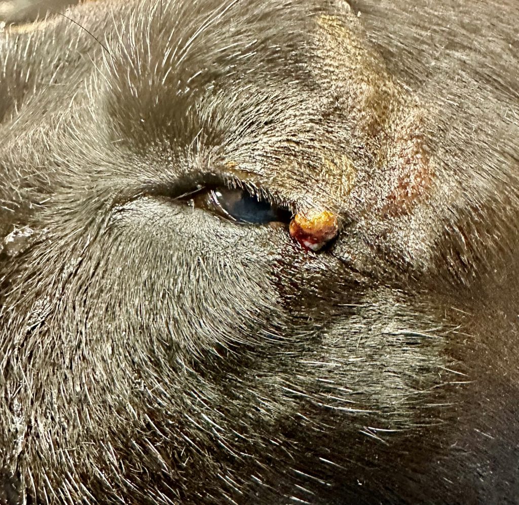
Meibomian gland adenomas (MGA) are another common abnormality that will show up in your exam rooms. Luckily, these are much more common than their adenocarcinoma cousins! These masses originate out of the cell lining of the meibomian glands and, in many cases, begin to rub against the cornea and cause irritation, or worse.
When the MGA are not interfering with the health of the cornea, I tend to have the owners monitor them until they begin to show signs of irritation for the patient. These masses can look wildly different per case, with some being smooth and nondescript, while others can be ulcerative, fragile, and bleed easily.
These masses almost always continue to grow on the eyelid margin, with the majority becoming a surgical candidate. From my experience, MGA occur much more often on older patients, which leads me to be more cautious with my anesthetic and surgical recommendations. Surgically, I remove a full thickness wedge of the eyelid, with margins several millimeters outside the width of the mass, as well as a few millimeters beyond what I can visualize the mass growing at the tip of the wedge.
I like to remove these masses with a CO2 surgical laser, but this can be performed with a scalpel, too. I close these incisions with a small dissolvable suture type, burying each individual suture and placing a small dab of surgical glue for added support. One recommendation when applying surgical glue to a site, such as this, is to pull up the surgical glue into a small syringe so that you can be more precise with the glue application. I recommend pathology on all these masses for our patients, so we know what to be watching for in the future.
A cherry on top
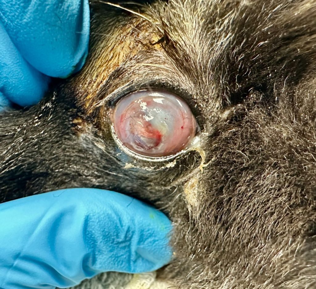
The dreaded “cherry eye.” If you have spent anytime within a veterinary hospital, you have seen one of these. As always, educating the client about how these occur is wise. The nictitating membrane is the third eyelid that raises diagonally in the inner corner of the eye in both dogs and cats. At its base is a tear gland referred to as the nictitans gland, which produces roughly 30 percent of the tears in the adjoining eye.
The prolapse of this gland can occur for several reasons, some inherited and some traumatic. When the tissue that keeps the gland in place at the base of the nictitating membrane is damaged or stretched, it allows the gland to push up out of its normal position and become visible. This mispositioning of the gland will diminish its ability to produce tears and, in some cases, can begin to irritate the patient’s cornea.
There are several surgical techniques to try and replace the nictitans gland to its normal positioning. The traditional tuck or tacking method and the imbrication of the tissues is the more common and highly recommended technique in saving this valuable tear gland. However, the suture needed for this procedure and the friability of the tissue involved lead these procedures to have a very high failure rate, and I would recommend explaining this to clients prior to preforming this surgery.
It will not and should not be surprising when these procedures need to be performed again for an adequate resolution.
Removing the nictitans gland is less desirable due to the fact it produces a sizable volume of the patient’s tearing. Any time the gland is damaged, prolapsed, or surgically replaced, I recommend preforming a Schirmer tear test and tracking tear production and changes. This should be considered important data to have over the duration of the tracking of this patient. I would also make the owner aware of the possibility of dry eye conditions later in life. When these conditions do begin to occur, the earlier we begin treatment, the better the overall outcomes. Additionally, educating our clients about the early signs of these ocular diseases can aid in prevention and treatment plans.
Common corneal impairments and lid abnormalities can and will be seen every day in a general veterinary practice. Taking the time with yearly continuing education to expand profession abilities in treating these ailments can make us invaluable to our patients and the practices we work within. If you have not been comfortable working on these in the past, take some time and work on these skills. With a little time invested, these are all procedures and treatments you can perform!
Cade M. Wilson, DVM, is a practicing veterinarian and a three-doctor mixed-animal practice owner in Ardmore, OK. Dr. Wilson has been practicing small animal medicine for the last 20 years and been a practice owner for the last
17 years.