Frequent itching, licking, and paw chewing are some of the most frustrating and common complaints of dog owners. These are among the top reasons for veterinary visits. Constant scratching and licking diminish the quality of life for the dog and their owner, impacting the human-animal bond.
Diagnosis
Canine atopic dermatitis (CAD) is a chronic inflammatory disease that is a diagnosis of exclusion. The pathogenesis of atopic dermatitis is complex. It is likely a defective skin barrier allows microbial adherence, penetration of allergenic proteins, and initiation of abnormal inflammatory and allergic responses.1
The evaluation of a pruritic dog requires a step-by-step approach2 and complicating factors must be ruled out.
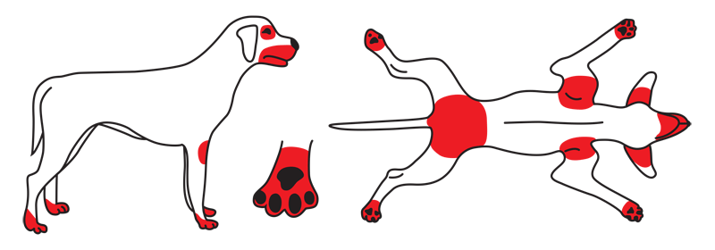
Favrot’s principles can help lead a clinician to a suspected diagnosis of CAD:4
- Initial itching without lesions in dogs younger than three years old
- An indoor lifestyle
- Affected feet and concave aspects of the pinnae
- Initial responsiveness to glucocorticoids administration3,4
However, it is vital to rule out other comorbidities and diseases that can present with pruritis and secondary skin infections. These may lessen the impact of treatment modalities, causing treatment failure and further frustration.
A detailed history of age of onset, seasonality, pruritis level, dietary trials, including treats and oral parasiticides, and previous treatments with their response should be collected. In addition, a minimum dermatological database consisting of skin and ear cytology, skin scrapings, and a DTM culture should be analyzed.
Once ectoparasitic diseases, superficial pyoderma due to bacteria and/or Malassezia, and food intolerance have been ruled out, the diagnosis of CAD can be made. Often, food can be one of the triggers for itching and inflammation in atopic dogs, leading to a multifactorial disease.6 Moreover, most CAD dogs tend to have a predisposition to secondary skin infections. Superficial bacterial pyoderma or Malassezia dermatitis can perpetuate and potentiate the disease.6
A critical aspect in managing both patient and the owner’s quality of life is reducing pruritis. Consider the use of antipruritic agents, such as glucocorticoids, oclacitinib, or lokivetmab.2 As these therapies may be less effective in the face of an active infection, appropriate diagnosis and treatment of secondary infections is critical before assessing response to antipruritic therapy.2
Disruption of skin barriers and CAD development
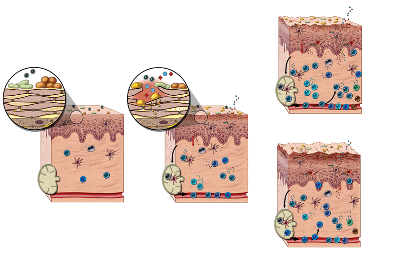
The skin’s protective function primarily relies on the outermost epidermal layer called the stratum corneum. This area is formed from tightly interlocked dead keratinocytes and surrounded by lipids and ceramides. The stratum corneum plays a vital role in protecting the skin from dehydration and reducing penetration of allergens.
In CAD, skin barrier disruptions are present, even in non-lesional skin. Impairment of the skin barrier is thought to enable the uptake of allergens, irritants, and microbes. In turn, these antigens trigger an innate response by inducing cytokines and Th2 cells which secrete interleukins responsible for the itch. These barrier defects are exacerbated by licking and scratching, which perpetuate infections and inflammatory cycles, causing more damage to the skin barrier (Figure 1).
Given CAD is an immune response to contact allergens, repairing the epidermal skin barrier is of utmost importance. The use of skin barrier-building materials, such as ceramides, lipids, ophytrium, and essential fatty acids (EFAs) that can repair the barrier should be used to minimize the immune response. As topical therapy directly treats the affected skin, adding this modality may reduce or potentially eliminate the need for systemic medications.6
Topical therapy
There are many topical therapies to choose from, including shampoos, mousses, sprays, and spot-on treatments. The act of bathing removes environmental allergens and can remove exudate and crusting. Several products contain ingredients thought to improve the skin barrier and reduce pruritis:
-
- Ophytrium is a purified natural ingredient extracted from the tuberous root of Ophiopogon japonicus. It has been shown to improve the mechanical, microbial, and immunological components of the skin barrier. In addition, it has been demonstrated to reduce the adhesion and biofilm formation of S. pseudintermedius on the surface of reconstructed canine epidermis.8 When applied to stressed reconstructed human epidermis, ophytrium allowed the tissue to recover to normal morphology by reducing transepidermal water loss and reducing proinflammatory cytokines.9
- Ceramides are key components of the extracellular lipid matrix in the stratum corneum. A significant decrease in the concentration and metabolism of ceramides was found in some CAD-afflicted dogs.10 Several products contain ceramides or pro-ceramides, such as phytosphingosine. In addition to moisturizing the skin barrier, they may also be anti-inflammatory and anti-pruritic.
- Combination products that contain a lipid complex, phytoceramides, and essential oils have been shown to help restore the ultrastructural lipid abnormalities and improve the severity of dermatitis in a small number of dogs with CAD.6,11
- Another topical formulation containing sphingomyelin-rich sphingolipids (a precursor of ceramides), plus glycosaminoglycans (GAGs) and hyaluronic acid, has been shown to improve the skin barrier and reduce clinical signs associated with CAD.
In one study, house dust mite allergic atopic beagles were challenged with allergen twice weekly. The treatment group showed a significantly lower canine atopic dermatitis extent and severity index (CADESI) score after one week, and a significantly lower pruritis visual analog score (PVAS) after eight weeks.11 A collar was also used, which showed improvement in CADESI-4 and PVAS scores in 12 client-owned dogs.12
The emergence of resistant bacteria and the need for judicious use of systemic antimicrobials makes topical therapy the recommended treatment for superficial pyoderma. In fact, topical therapy can be the sole treatment and has been found to be as effective as systemic antimicrobials or antifungals.14,15 For dogs with mild secondary bacterial or fungal infections, a shampoo with the active ingredients of chlorhexidine +/- miconazole has been shown to be an effective treatment.13
When treating with medicated shampoos, the frequency and duration of exposure will depend on each patient. Most guidelines call for shampooing two to three times per week with a minimum of 10 minutes of contact time before rinsing with cool water. Treatment should continue for a minimum of seven days past clinical resolution of signs or a negative skin cytology.
While using medicated shampoos is as efficacious as systemic therapy, compliance can be an issue. Clients typically have concerns about the frequency of bathing and may skip baths or treatments. Newer studies combining shampoo with a medicated mousse in lieu of frequent bathing may help improve compliance and speed clinical resolution.14,15
Dietary management
Newer innovations in dietary management are also valuable in building the skin barrier from within. Dogs with CAD have impaired intracellular lipid lamella of the stratum corneum and lower ceramide levels. Evidence suggests oral supplementation with polyunsaturated fatty acids (PUFAs) can reduce pruritis and lower the dosages of cyclosporine and glucocorticoids required to control clinical signs.16,17
PUFAs are thought to combat CAD by enhancing epidermal barrier function, reducing inflammatory cell activation, and altering eicosanoid production. The addition of antioxidants, PUFAs, polyphenols, and omega-3 fatty acids can reduce the clinical signs of CAD and are incorporated in many commercial diets formulated for CAD.
Nutraceuticals
Palmitoylethanolamide (PEA-um) is a naturally occurring bioactive lipid, produced on-demand by damage-exposed cells. According to one study, PEA-um appeared to be effective and safe in reducing pruritus and skin lesions, and in improving quality of life in dogs with moderate atopic dermatitis and pruritus.18
Extracts derived from hardy kiwi (Actinidia arguta) fruit have been shown to change CADESI-03 scores in atopic dogs when used with steroids, with those lower CADESI scores maintained after steroids have been tapered off. The fruit contains phenolic acids and flavonoids that may help to regulate the allergic response and inflammatory mediators.19
Oral supplementation with essential fatty acids (EFAs) has been shown to improve coat quality and skin barrier function in many CAD patients. A randomized controlled trial established that the daily administration of an EFA supplement allowed for a reduced dosage of oral prednisolone needed to control pruritis in dogs with CAD and may have a steroid sparing effect.20
In summary
Unfortunately, CAD cannot be cured and requires lifelong management. By improving the skin barrier, frequent flares can be minimized and allow a more comfortable existence for both clients and their dogs.
When managing CAD, it is important to determine the right combination of therapies for each individual dog that will provide a safe, effective, and affordable management strategy the owner can execute.6
Klarissa Ligon, DVM is a veterinary services manager for Ceva Animal Health. Dr. Ligon received her doctorate in veterinary medicine from Virginia Maryland Regional College of Veterinary Medicine, with an interest in small animal medicine and dermatology. Dr. Ligon has worked in the veterinary management and consolidation space. She is a Fear Free certified professional and has special interests in dermatology, behavior, and Chinese Shar Pei health disorders.
References
-
- Update on pathogenesis, diagnosis, and treatment of atopic dermatitis in dogs. Nuttall, et al. June 1, 2019, JAVMA, p. Vol 254 No. 11; 1291.
- 2023 AAHA Management of Allergic Skin Diseases in Dogs and Cats Guidelines. Miller J, Simpson A, Bloom P, et al. s.l. : Journal Am Anim Hosp Assoc, 2023, Vols. 59:255-284.
- Canine Atopic dermatitis: detailed guidelines for diagnosis and allergen identification. Hensel P, Santoro D, Favrot C, et al. 2015, BMC Vet Res, pp. 11:196-209.
- A prospective study on the clinical features of chronic canine atopic dermatitis and its diagnosis. Favrot C, steffan J, et al. s.l. : Veterinary Dermatology, 2010, Vol. 21.
- Review: Clinical and histological manifestations of canine atopic dermatitis. Bizikova, Petra, et al. 2, 2015, Veterinary Dermatology, Vol. 26, p. 79.
- Current Knowledge on Canine Atopic Dermatitis: Pathogenesis and Treatment. Outerbridge, et al. 2021 November, Adv Small Anim Care, pp. 2:101-115.
- Atopic Dermatitis. Weidinger S, Novak N. s.l. : The Lancet, 2016, Vols. 387: 1109-22.
- Effect of the ingredient A97614A1 on the adhesion and biofilm formation of Staphylococcus pseudintermedius in a model of reconstructed canine epidermis. Ollivier E, Zermirline C, Marchand, L, Closs B. s.l. : BSAVA Congress, 2019.
- Efficacy of the ingredient A97614A1 in a model of reconstructed human epidermis stressed by cytokines. Ollivier E, Zemirline C, Amalric N, et al. 2019 BSAVA Congress.
- Decreased concentration and enhanced metabolism of sphingosine-1-phosphate in lesional skin of dogs with atopic dermatitis: distrubed sphingosine-1-phosphate homeostasis in atopic dermatitis. Baumer W, Rossback K, Mischke R, et al. s.l. : Journal of Investigative Dermatology, 2011, Vols. 131(1):266-268.
- Topical treatment with Sphingolipids and Glycosaminoglycans for canine atopic dermatitis. Marsella, et al. 2020, BMC Veterinary Research, p. 16:92.
- Sphingomyelin-Rich Lipid Extract Collar for Canine Atopic Dermatitis. Segarra, et al. 2023, Veterinary Sciences, pp. 10,389.
- Biology, diagnosis and treatment of Malassezia dermatitis in dogs and cats – clinical consensus guidelines. Bond R, Morris, DO, Guillot J, et al. s.l. : Veterinary Dermatology, 2020, Vols. 31(1); 27-e4.
- Performance of two protocols combining Chlorhexadine digluconate 3% and Ophytrium-based shampoo and mousse applications in the management of bacterial overgrowth in dogs. Gatellet M, Bruet V, Cadot P-M, et al. Milan, Italy : s.n., 2020. 14th UNISVET National Congress.
- Performance of a combination of Ophytrium-containing shampoo and mousse applications in the management of sensitive skin in dogs: intermediate results of a field study. Gatellet M, Keteman R, Blondel T, et al. Milan, Italy : s.n., 2020. 14th UNISVET National Congress.
- Non-controlled, open-label clinical trial to assess the effectiveness of a dietetic food on pruritus and dermatologic scoring in atopic dogs. Witzel-Rollins, Angela, et al. 1, 2019, BMC Veterinary Research, Vol. 15, p. 220.
- Randomized, double-blind, placebo-controlled clinical trial measuring the effect of a dietetic food on dermatologic scoring and pruritus in dogs with atopic dermatitis. de Santiago, et al. 2021, BMC Veterinary Research, p. 17:354.
- Efficacy of ultra‐micronized palmitoylethanolamide in canine atopic dermatitis: an open‐label multi‐centre study. Noli, Chiara, et al. 6, 2015, Veterinary Dermatology, Vol. 26, p. 432.
- A randomized, double-blind, placebo-controlled study to evaluate the effect of EFF1001, an Actinidia arguta (hardy kiwi) preparation, on CADESI score and pruritus in dogs with mild to moderate atopic dermatitis. Marsella, Rosanna, et al. 1, 2010, Veterinary Dermatology, Vol. 21, pp. 50-57.
- A randomized, controlled study to evaluate the steroid sparing effect on essential fatty acid supplementation in the treatment of canine atopic dermatitis. Saevik BK, Bergvall K, Hom B, et al. s.l.: Veterinary Dermatology, 2004, Vols. 15(3):137-145.
- Current evidence of skin barrier dysfunction in human and canine atopic dermatitis. Marsella, Rosanna, Olivry, Thierry and Carlotti, Didier‐Noël. 3, 2011, Veterinary Dermatology, Vol. 22, pp. 239-248.
- Skin Barrier Reinforcement Effect Assessment of a Spot-on Based on Natural Ingredients in a Dog Model of Tape Stripping. Idee A, Mosca M, Pin Didier. s.l. : Veterinary Sciences, 2022, Vols. 9, 390.