As in domestic small mammals, radiography is an essential diagnostic tool for reptiles, birds, and small companion mammals, including ferrets, rabbits, and rodents. These species can present several novel challenges to veterinary practitioners due to the unique body shapes and sizes, as well as the need for sedation to facilitate proper positioning for many exotic companion animals.
Reptiles
Restraint
Many reptile patients can be radiographed with manual restrain alone, without the need for sedation or anesthesia. The exception to this is snakes—even the most tractable snake will often not remain still (and completely straight) in any position for long enough to obtain two views of their whole body. Even if placed in an acrylic clear tube, snakes often are not completely straight if they are fully awake, which can complicate radiographic interpretation. The snake should be anesthetized or heavily sedated (such as with intramuscular alfaxalone). Two views (lateral and dorsoventral) of the whole body (including head and tail) should be obtained with the snake’s body fully extended.
For most chelonians, no chemical or manual restraint is necessary for whole-body views. However, if radiographs of the limbs or skull are needed, then heavy sedation or general anesthesia is often required to extend the skull and/or limbs from the shell. Intramuscular combinations of ketamine, dexmedetomidine, +/- midazolam are the most common protocols in my practice in this taxa.
Lizards also rarely require restraint for routine whole-body radiographs, but if needed, intramuscular combinations of ketamine, dexmedetomidine, +/- midazolam or alternatively intramuscular alfaxalone alone (in smaller lizards) can be used.
Positioning
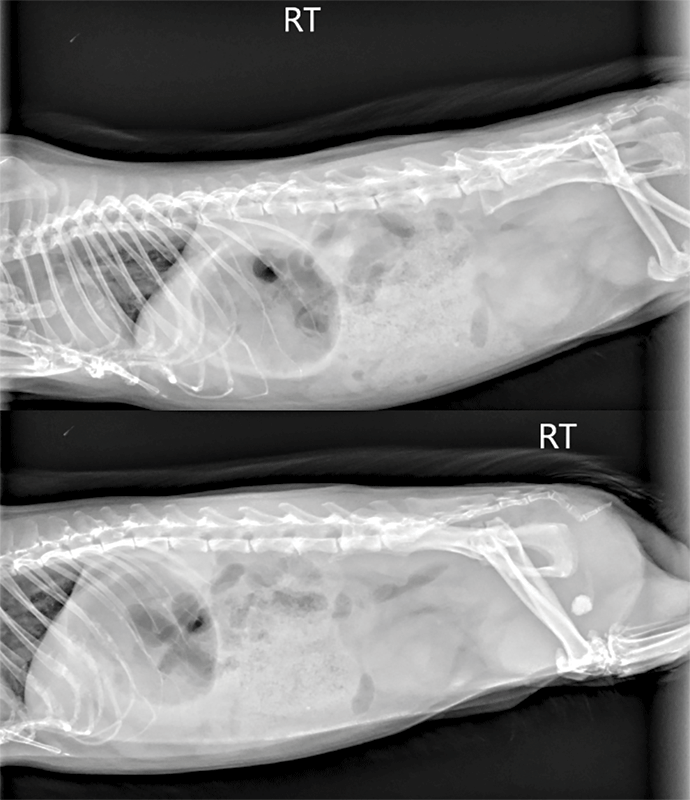
- Lizards. It is standard to take whole-body views as most reptiles do not have a separate thorax and abdomen, but rather one continuous cavity (coelom). For lizards, dorsoventral and horizontal beam lateral (right or left) views are recommended. A horizontal beam is highly preferred due to displacement of coelomic organs that occurs when a lizard (lacking a diaphragm) is placed on their side. This is especially important for dorsoventrally compressed lizards, such as bearded dragons (Figure 1). The exception to this would be laterally compressed lizards, such as chameleons, which tend to have less internal organ shift when placed in lateral recumbency.
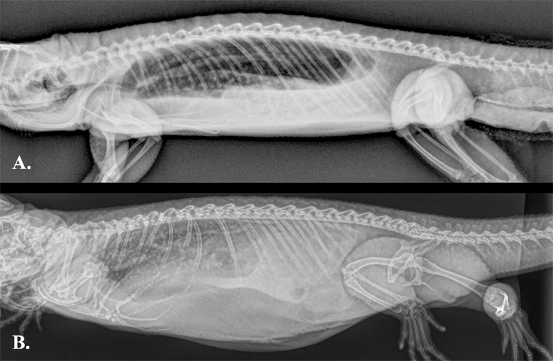
Figure 1. Two whole-body lateral views of an adult bearded dragon. Note the difference in radiographic appearance, particularly of the lung fields, between the image obtained with a horizontal beam (A) compared with a vertical beam (B). Photo courtesy Dr. Olivia Petritz When positioning lizards for a horizontal-beam lateral view, it is important to ensure the limbs are extended downward to limit superimposition on the coelom. This is especially important for the forelimbs as the trachea, heart, and great vessels lie between the pectoral girdle in most lizard species. This can be accomplished by setting the lizard on a piece of foam or other non-radiopaque device narrow enough to allow the limbs to hang ventrally. Most lizards will tolerate this position for brief periods of time without any additional manual or chemical restraint.
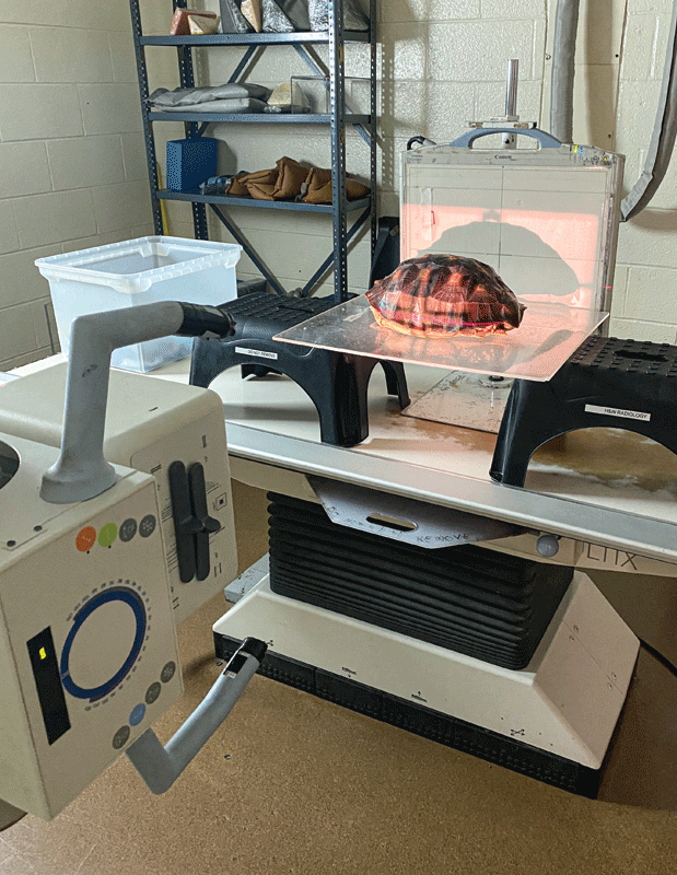
- Chelonians. Three standard radiographic views should be obtained for chelonians: dorsoventral, horizontal beam lateral (right, left, or both), and rostro-caudal views.
The rostro-caudal view is important to facilitate interpretation of each lung field independently without superimposition. Ideally, the chelonian patient would be positioned on an elevated platform (plastic tub/bowl) to allow all four limbs to hang ventrally. However, in my clinical experience, it is uncommon for a non-sedated chelonian patient to tolerate this position, unlike in lizards.
Similar to lizards, the use of a horizontal beam prevents the shifting of coelomic organs which occurs when a chelonian is restrained in lateral recumbency vertically, in the case of the rostro-caudal view (Figure 2).
- Snakes. Two views (lateral and dorsoventral) of the whole body (including head and tail) should be obtained with the snake’s body fully extended. If the entire snake does not fit on a single view, then multiple views should be obtained, with numbered markers to help delineate and organize the sections and digital images.
A lateral view is superior to the dorsoventral view to examine most coelomic structures. Using a horizontal beam is also preferred for snakes, but there is less artifact of coelomic organ displacement when using a vertical beam in this taxa compared with other reptiles.
A dorsoventral view of a coiled snake can lead to distortion of internal organs and asymmetry of spine and ribs; therefore, proper positioning is highly recommended to facilitate complete interpretation.
Interpretation
Reptile radiology is unfortunately characterized by poor serosal contrast due to the close proximity of internal organs, lack of distributed internal fat (as most is stored in organized coelomic fat bodies), lack of clearly demarcated thorax and abdomen, and image degradation due to superimposed exoskeleton (plastron and carapace) or cutaneous scales (which can be mineralized in the case of osteoderms).
Due to the density of larger chelonians, small animal radiology machines are often not powerful enough to create quality radiographic images, even if the turtle or tortoise can still physically fit on a large digital plate. In addition, their intracoelomic detail on survey radiographs is often quite poor even with appropriate radiographic technique. Typically, only the trachea, lungs, heart, gastrointestinal tract, and sometimes the liver are easily identified on survey radiographs in snakes and lizards.
Further, due to the anatomical differences between reptilian and mammalian lungs, most of the radiographic pulmonary patterns (alveolar, bronchial, etc.) as described for mammals are not applicable to reptiles.
Ultimately, additional imaging modalities, such as coelomic ultrasound and/or a CT scan, are often needed to further evaluate most intracoelmic organs in reptiles. The use of gastrointestinal contrast can be helpful to further delineate the gastrointestinal tract and evaluate transit time in most reptile patients—there are multiple publications reporting normal values for this technique in several lizard, snake, and chelonian species.1-6
Birds
Restraint
Chemical restraint almost always requires for proper radiographic positioning of avian species. Raptors are an exception to this, and often will be tractable for radiographs with hooding in my experience. Sedation with midazolam and butorphanol, either IM or intranasal, are the most common drug protocols used in my practice. The midazolam can be reversed with 0.05-0.1mg/kg flumazenil IM/IN, so this can be an outpatient procedure.
If the bird requires additional sedation or needs to hold perfectly still, then general anesthesia will likely be required in addition to parenteral sedation drugs.
Positioning
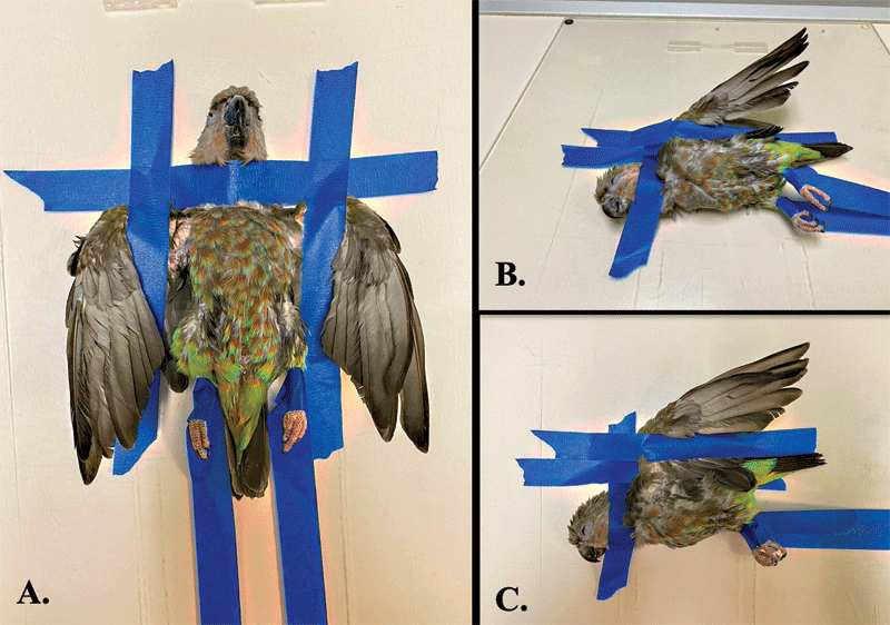
It is standard to take whole-body views because, similar to reptiles, birds do not have a separate thorax and abdomen, but rather one coelomic cavity. For the ventrodorsal view, the bird should be in dorsal recumbency, with wings extended equally and taped to the plate, while the pelvic limbs are extended as far caudal as possible and taped. The keel should be superimposed upon the spine.
The lateral view requires the bird in right lateral recumbency, with both wings extended dorsally above the spine and held in place with tape. The pelvic limbs should again be extended as far caudally as possible to minimize superimposition with the coelomic structures. The coracoids and coxofemoral joints should be superimposed. Either paper tape or painting tape should be used as other types (white tape or waterproof white tape) can damage or remove their feathers. (Figure 3). Commercial plastic restraint boards are also available to assist with positioning.
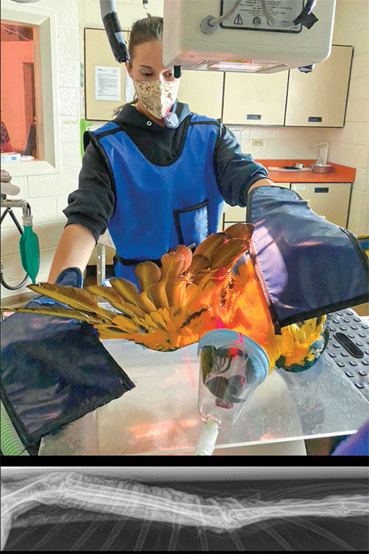
To evaluate the wings, a collimated lateral and the orthogonal caudo-cranial view is required. The latter of which requires the wing to be extended laterally and pronated, with the primary flight feathers extending upwards and perpendicular to the plate (Figure 4). A new imaging technique has also been described to examine the pectoral girdle in raptor species.7 It is also highly recommended to develop a technique chart for a variety of birds (by size or by weight) specific for each radiographic machine. The technique is typically the same between the lateral to the ventrodorsal view on the same patient.
Interpretation
As in other species, radiographic technique and positioning are extremely important for complete and accurate interpretation of avian radiographs. There are several reference textbooks that have published normal radiographs from a variety of avian species; however, most of these have nondigital images, which can be challenging to compare with digital radiographs.
In addition, there are several articles that have published normal radiographic measurements for different organs, mainly of psittacines, including proventricular size via the proventricular: keel ratio and cardiac size via cardiac: thoracic ratios. A review article summarizes these published studies in psittacines.8
Small mammals
Restraint
Chemical restraint is often required for appropriate positioning of these species, and sedation, rather than general anesthesia, is typically recommended. Some animals are tolerant of brief manual restraint, but complications, such as iatrogenic trauma (i.e. vertebral fractures in rabbits) and undue stress in often compromised patients can occur.
Intramuscular combinations of midazolam and butorphanol +/- ketamine or alfaxalone are the most common sedation protocols used in my practice. Midazolam can be reversed with flumazenil (IM or IV). Mask or chamber induction with inhalant anesthetics without premedication is not recommended, as rabbits will experience profound hypotension secondary to even brief exposure to inhalant anesthetics.
Positioning
Standard views to evaluate the thorax and abdomen include a laterolateral (either right, left, or both) and ventrodorsal or dorsoventral projections. Whole-body radiographs, rather than separate thoracic and abdominal, are often performed due to small patient size; however, in larger rabbit patients, separation of abdomen and thoracic views is easily accomplished.
Positioning patients for these films is identical to positioning for domestic dogs and cats. Care should be taken to fully extend the forelimbs and hindlimbs to limit superimposition with the thorax/abdomen. If urolithiasis (common in guinea pigs) is suspected, lateral views should include the entire aspect of the caudal abdomen and tail to ensure the whole urethra can be evaluated (Figure 5). In addition, it is often helpful to take several views with hindlimbs held cranially and then caudally.
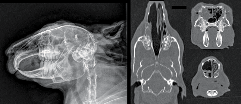
Standard views to evaluate the skull include a laterolateral (either right, left, or ideally both), right lateral oblique, left lateral oblique, and ventrodorsal or dorsoventral views. Additional views, including a rostro-caudal, and open vs. closed mouth projections, can be obtained; however, that is outside of the scope of this review.
Proper positioning is key for correct interpretation of skull radiographs, and this almost always requires heavy sedation of the patient. A straight laterolateral view will have both bullae superimposed. Minimal rotation of the skull is required for the oblique views, which function to highlight the ventral aspect of both mandibles. Skull radiographs have limited usefulness to evaluate the soft tissues of the head and the nasal passages. Certain disease processes, such as rhinitis, often are not visible radiographically unless they are severe and bilateral.
Since many rabbits and rodents have concurrent dental and upper respiratory tract disease, an imaging modality that can evaluate both bone and soft tissue is preferred, such as a computed tomography (CT) (Figure 6). CT scans are extremely valuable and are often performed with a conscious or sedated patient, rather than utilizing general anesthesia. With many of the newer CT machines, the scanning time is significantly reduced (i.e. five to 10 minutes). Normal CT anatomy of the rabbit skull has been previously published for reference in clinical cases.9
Interpretation
Prior to interpretation, evaluation of the radiographic technique and positioning is extremely important, especially if it is an outpatient procedure. As in domestic dogs and cats, a systematic approach for radiographic interpretation in rabbits and rodents is recommended. I use a systems approach, in the following order: musculoskeletal, pulmonary, cardiac, hepatic, gastrointestinal, and finally the urinary/reproductive systems. The rabbit thorax, especially the cranial thorax, is challenging to interpret due to small patient size, and persistence of the thymus gland into adulthood. Due to the complex nature of the gastrointestinal tract, this, too, is a challenging area to interpret for most rabbits and herbivorous rodents. The lower gastrointestinal tract (small intestine, large cecum, and most sections of the colon) are superimposed on one another and does not have the spatial separation as is seen in domestic dogs and cats.
Skull films can be evaluated in the following order: musculoskeletal, bullae, nasal passages, incisors, maxillary premolars-molars, mandibular premolars-molars, intraoral occlusion, and then soft tissue structures of the head. A series of reference lines (Figure 1) were developed by researchers to help clinicians objectively interpret skull radiographs in rabbits, guinea pigs, and chinchillas.10 In a more recent paper,11 these same researchers compared the use of these skull radiographic reference lines across different rabbit breeds—they determined that in rabbits with a large palatal angle (those breeds with shortened skulls) these reference lines could not be applied in the same manner across breeds.
Olivia A. Petritz, DVM, DACZM, graduated from Purdue University, and then completed several internships and a residency in the field of zoo and exotic animal medicine. Dr. Petritz became a diplomate in the American College of Zoological Medicine in 2013 and specializes in zoological companion animals (exotic pets). Petritz started an exotics service at a specialty hospital in Los Angeles following her residency, and currently is an associate professor of Avian and exotic animal medicine at North Carolina State University.
References
- Long, Charles Tyler, et al. “Comparison of Gastrografin to Barium Sulfate as a gastrointestinal contrast agent in Red‐eared slider turtles (trachemys Scripta Elegans).” Veterinary radiology & ultrasound 51.1 (2010): 42-47.
- Smith, D., H. Dobson, and E. Spence. “Gastrointestinal studies in the green iguana: technique and reference values.” Veterinary Radiology & Ultrasound 42.6 (2001): 515-520.
- Grosset, Claire, et al. “Radiographic anatomy and barium sulfate contrast transit time of the gastrointestinal tract of bearded dragons (Pogona vitticeps).” Veterinary Radiology & Ultrasound 55.3 (2014): 241-250.
- Mathes, Karina A., et al. “Specific anatomy and radiographic illustration of the digestive tract and transit time of two orally administered contrast media in Inland bearded dragons (Pogona vitticeps).” Plos one 14.8 (2019): e0221050.
- Houck, Emma L., et al. “Radiographic anatomy and barium sulfate contrast study of the gastrointestinal tract of eastern box turtles (Terrapene carolina carolina).” Veterinary Radiology & Ultrasound 60.5 (2019): 473-484.
- Banzato, Tommaso, et al. “Development of a technique for contrast radiographic examination of the gastrointestinal tract in ball pythons (Python regius).” American journal of veterinary research 73.7 (2012): 996-1001.
- Visser, Marike, et al. “Use of a caudoventral-craniodorsal oblique radiographic view made at 45° to the frontal plane to evaluate the pectoral girdle in raptors.” Journal of the American Veterinary Medical Association 247.9 (2015): 1037-1041
- Geerinckx, Lise, et al. “Literature Review of Radiographic Measurements of Internal Organs in Psittaciformes.” Journal of exotic pet medicine 28 (2019): 60-68.
- Van Caelenberg, Annemie I., et al. “Computed tomography and cross-sectional anatomy of the head in healthy rabbits.” American journal of veterinary research 71.3 (2010): 293-303.
- Boehmer, E., and D. Crossley. “Objective interpretation of dental disease in rabbits, guinea pigs and chinchillas.” Tierärztliche Praxis 37 (2009): 250-260.
- Böhmer, Christine, and Estella Böhmer. “Skull shape diversity in pet rabbits and the applicability of anatomical reference lines for objective interpretation of dental disease.” Veterinary sciences 7.4 (2020): 182.
Additional reading:
- Schmidt, Lauren, Nicola Di Girolamo, and Paolo Selleri. “Diagnostic Imaging of the Reptile Urinary System.” Veterinary Clinics: Exotic Animal Practice 23.1 (2020): 131-149
- Gumpenberger, Michaela. “Diagnostic Imaging of the Respiratory Tract of the Reptile Patient.” Veterinary Clinics: Exotic Animal Practice 24.2 (2021): 293-320.
- Silverman, Sam, and Lisa Tell. Radiology of rodents, rabbits and ferrets-E-book: an atlas of normal anatomy and positioning. Elsevier Health Sciences, 2004.
- Capello, Vittorio, and Angela M. Lennox. Clinical radiology of exotic companion mammals.
- John Wiley & Sons, 2013. Silverman, Sam, and Lisa A. Tell. Radiology of birds: an atlas of normal anatomy and positioning. Elsevier Health Sciences, 2010.