Understanding a species’ standard health condition in the wild is critical to informing care. However, doing so can be a challenge, especially as a radiologist in a specialty area of care. Most diagnostic X-ray systems cannot be transported to or perform in the rugged conditions of an animal’s habitat, but bridging this knowledge gap can be the key to elevating the well-being of a species in both the wild and in human care.
The endangered Matschie’s tree kangaroo, endemic only to the Huan Peninsula of Papua New Guinea, is a powerful example of this principle. Over the past 25 years, the Seattle-based Woodland Park Zoo’s Tree Kangaroo Conservation Program (TKCP) has worked with local landowners and Papua New Guinea’s government to conserve this species, which is endangered due to over-hunting and loss of habitat. TKCP has helped the government of Papua New Guinea establish the YUS Conservation Area, named for the Yopno, Uruwa, and Som Rivers that run through the area, as the first nationally recognized community-managed conservation area in the country’s history.
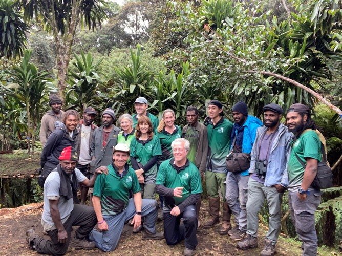
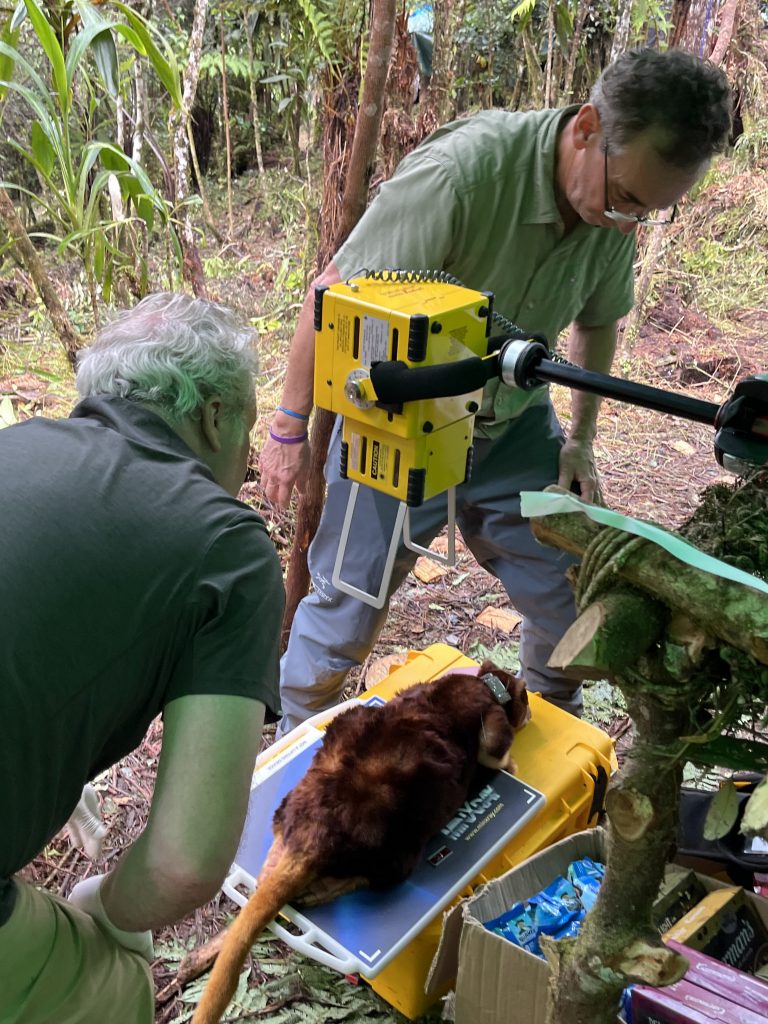
I recently traveled with wildlife biologists and veterinarians to a TKCP tree kangaroo research base located in the YUS Conservation Area at an elevation of 10,000 feet. The team included TKCP scientists, led by director Lisa Dabek, PhD, as well as zoo wildlife veterinarians, Carol Esson, MVSc, PhD, Marisa Bezjian, DVM, and Tim Storms, DVM. We worked with local tracking specialists to humanely capture tree kangaroos, which were carefully anesthetized for a complete physical examination. This exam included measuring the size and shape of the body, inspecting pouches on females, analyzing acquired blood samples, and obtaining radiographs and screening ultrasounds of the animals. Before each animal was released back to the tree from which they were taken, we also microchipped and GPS-collared them for future data collection.
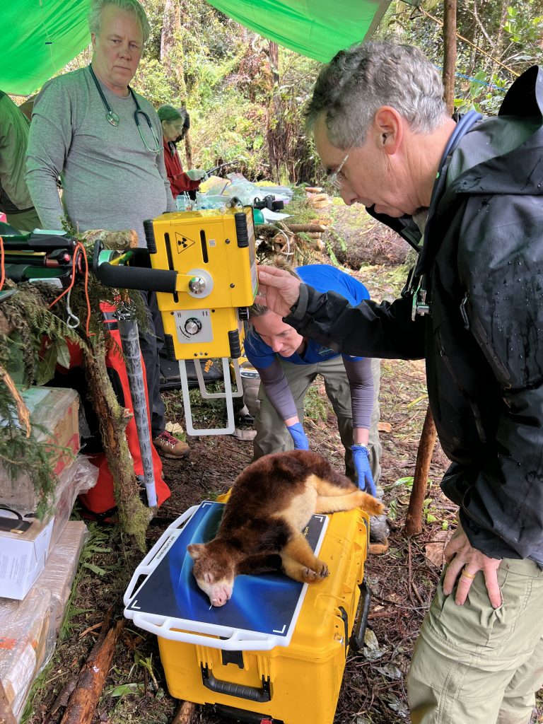
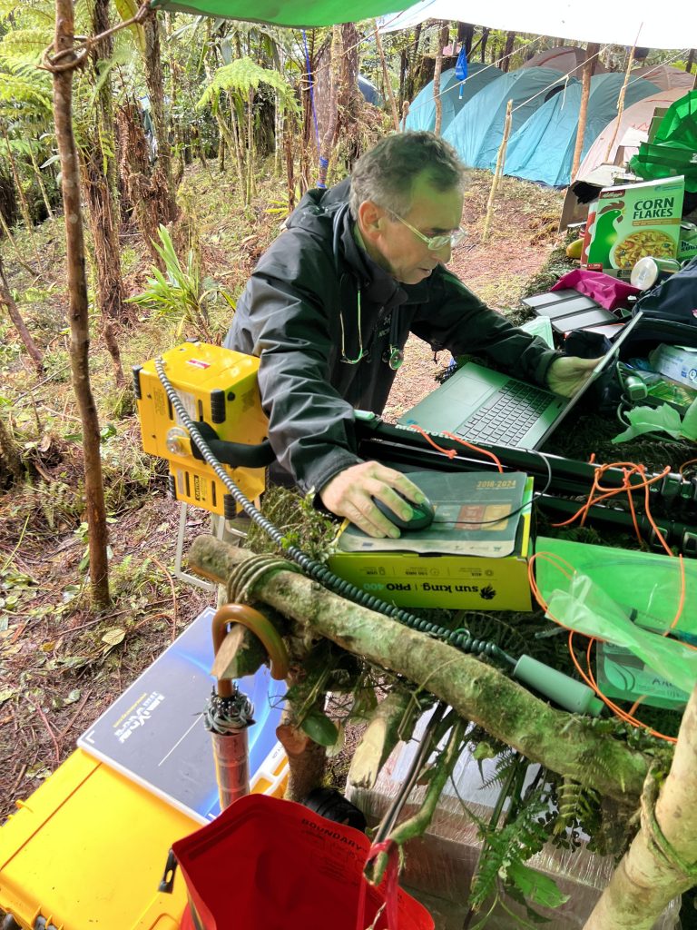
Despite the rain, elevation, and lack of power, I was able to use an ultra-portable, battery-powered digital radiography system to capture high-quality diagnostic images. The batteries were charged using solar panels, allowing us to use the X-ray system for the entire 10 days we were at the research center.
The images we obtained helped us to establish a baseline for the Matschie’s tree kangaroo and assess for differences between animals in human care and the wild. With this information, we will be able to improve our approach to caring for both populations and continue our work towards recovering this endangered species.
MAC infections
During the most recent trips to the research camp, one of our primary goals was to test the hypothesis that the mycobacterium avium complex (MAC) infections found in Matschie’s tree kangaroos in human care are related to their environment and therefore would not be found in wild animals.
MAC is ubiquitous, present in soil and water, and causes infections in humans and various animals. It rarely causes an infection in humans unless the person is severely immunocompromised. However, it can also infect an animal with a weak immune system. Marsupials tend to have less robust immune capacities, and initial research comparing zoo animals to their wild counterparts has also found the former may have weaker immune systems. This combination of factors theoretically may cause Matschie’s tree kangaroos in human care to be more susceptible to MAC infections.
Though the disease is often terminal in tree kangaroos, MAC infections are slow to develop, with most infected animals surviving for many months after diagnosis. This made the team reasonably certain if the disease were present in wild animals, signs would show in the tree kangaroos we analyzed.
Using an ultra-portable, digital X-ray system, we imaged 14 wild Matschie’s tree kangaroos in the YUS Conservation Area. None have exhibited signs of MAC, such as lung nodules or calcifications in lungs, mediastinum, liver or spleen, as well as no radiographic evidence of MAC osteomyelitis or septic arthritis. Blood and gastric aspirates collected from the animals are still being analyzed to confirm that none of the mycobacteria is present.
While more assessment is needed the lack of any MAC-infected wild tree kangaroos currently known to us supports the hypothesis tree kangaroos in human care are susceptible to MAC infection due to an environmental cause. That cause is not proven at this point, but potential etiologies can include long-term exposure to nonnatural habitat or nutritional deficiencies, either of which could lead to immunosuppression.
Skeletal injuries
Musculoskeletal trauma can happen in both wild and zoo environments. While relatively uncommon, Matschie’s tree kangaroos in zoos can fall awkwardly and injure themselves.
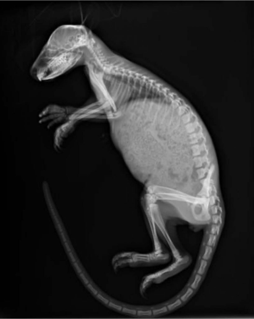
In the wild, the behaviors of the Matschie’s tree kangaroo would logically make them more susceptible to skeletal injuries to the limbs and tail. One of the animal’s natural predators is the New Guinea harpy eagle. To avoid capture by this predator, a tree kangaroo will jump from its perch in a tree to the ground when it senses an eagle approaching.
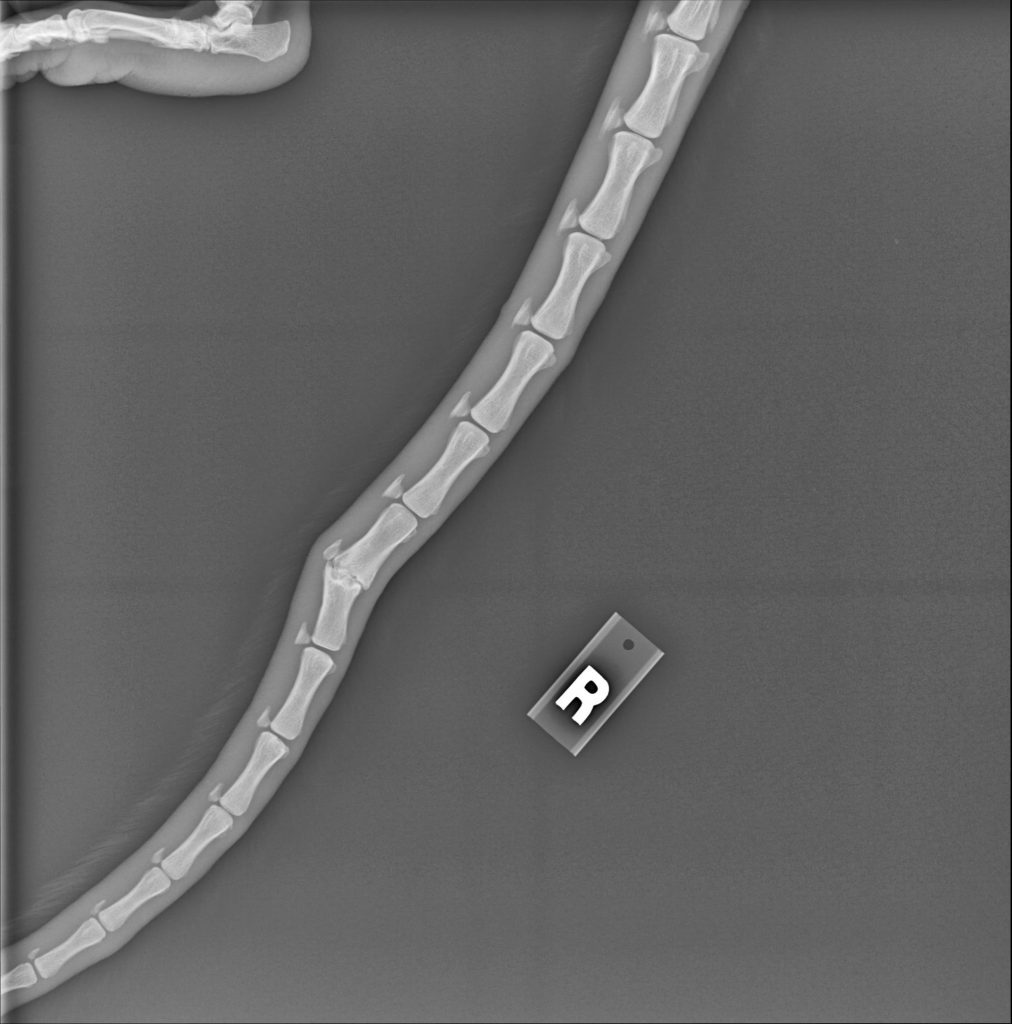
Trees grow much taller in the rainforests of Papua New Guinea than in a zoo enclosure, and this can lead to a drop of 75-100 feet for the animal. We saw evidence of related trauma in the kangaroos we radiographed, including old injuries to tail vertebrae and old, healed rib fractures. However, we saw less evidence of skeletal trauma than expected. Given their frequent free falls, the animals were remarkably healthy.
With this information, we can work to mimic the Matschie’s tree kangaroo’s natural setting to minimize trauma and preserve bone health.
One Health
Recognizing there is a close, interdependent relationship among the health of humans, wildlife, and the environment, a cornerstone of TKCP’s approach to conservation is the One Health model. Throughout its history, TKCP has worked to support this mission by developing local knowledge, capacity, and leadership efforts to maintain the health of the community.
Over 80 percent of Papua New Guinea’s population lives in remote areas, meaning access to medical equipment and services requires a multi-day walk. After our 10 days in the rainforest, we spent an additional 10 days in a remote village, providing healthcare education, diagnostic technology and specialty care that would otherwise be inaccessible.
I was joined by several other specialists including a nutritionist and health education trainers from the region, along with a physical therapist, emergency room doctor, OB-GYN, physician and an internist. Over 400 people traveled from adjacent villages to attend our healthcare education seminars, and we were able to see approximately 200 patients, some of whom walked for days to be seen. We were once again able to keep our equipment charged for the duration of our visit by using solar panels, and the villagers built a temporary isolated room for the X-ray equipment since lead shielding was not possible.
Using the battery-powered ultra-portable radiography equipment and portable ultrasound, I was able to diagnose cancers, infections and various musculoskeletal conditions, as well as screen for common diseases that have a high burden in the area, including emphysema and tuberculosis.
“We were thrilled to loan a machine to such an important cause and see the impact it had on both tree kangaroo conservation and healthcare in the local village,” says Jeanne Walter, vice president of marketing and sales for MinXray, the company that provided the X-ray system for the trip. “Our equipment was designed to be adaptable to any species, environment and condition to ensure that the important healthcare resource of radiographic imaging can be accessible anywhere.”
In addition, our partnerships with a wide network of stakeholders, including the Morobe Provincial Health authority, allow us to support broader One Health initiatives across Papua New Guinea. Peer education programs throughout the YUS landscape have helped promote awareness of One Health and address primary health concerns, such as nutrition, water quality and access, hygiene and sanitation, and respiratory illness through preventative measures and nature-based solutions.
Going forward, TKCP is looking to continue learning more, helping the Matschie’s tree kangaroo population to thrive, and working with the Morobe Provincial Health Authority to further integrate priorities for human health into the community.
Robert Liddell, MD, is a Seattle-based semi-retired diagnostic radiologist. Since his residency days at the University of Washington, Dr. Liddell has volunteered as a radiologist consultant to the veterinary staff at the Woodland Park Zoo in Seattle, Wash., providing ultrasound, CT, and MRI expertise on a wide array of animals. Liddell also volunteers as a physician with the zoo’s Tree Kangaroo Conservation Program (TKCP), providing diagnostic imaging on the endangered Matschie’s tree kangaroo at a remote research camp in Papua New Guinea and working with other volunteer physicians to provide human medical care and education in villages of the country’s YUS Conservation Area.