Blood transfusions are used for various medical conditions to replace lost components of the blood. However, in veterinary medicine, blood products are not always readily available and can be cost-prohibitive. For this reason, autotransfusion can be a life-saving alternative to common blood products, as long as the emergency clinician knows and understands the clinical indications and possible complications of this procedure.
Hemorrhagic shock is commonly found in companion animals presenting to an emergency room. Trauma, anticoagulant rodenticide, and bleeding neoplasia represent common conditions that can be associated with severe hemorrhage.
During hemorrhage, a decrease in intravascular blood volume and an acute decrease in red blood cells can lead to a significant decrease in oxygen delivery to the peripheral tissue and cells, causing a state of shock.
The treatment goals during hemorrhagic shock are to replenish the missing intravascular volume and ensure adequate oxygen carrying capacity remains. The use of blood products, in addition to isotonic crystalloids, may, therefore, be vital in some hemorrhagic patients.
Benefits of autotransfusion
Autotransfusion is when an animal receives a transfusion of autologous blood (its own blood) instead of allogenic (different donor) blood. The transfusion of autologous red blood cells offers multiple advantages over the use of stored blood products.
First of all, by using the patient’s own blood, any concern with disease transmission or compatibility problems disappears. This can allow for quickly administering the autologous blood. The risk of transfusion reactions is also reduced.
Additionally, the blood is usually normothermic when collected from the pleural or peritoneal cavities, and rapid autotransfusion can, therefore, cause less hypothermia in the patient.
Lastly, most autotransfusions do not require anticoagulant and are therefore less likely to cause transfusion-associated citrate toxicity, which is caused by the binding of citrate to calcium and magnesium. When large quantities of stored anticoagulated blood are used, this can be associated with clinically relevant hypocalcemia and hypomagnesemia.
Before performing an autotransfusion
Choosing the right patient for autotransfusion is important for the success of the procedure. The ideal patient for autotransfusion is one that remains unstable despite standard isotonic crystalloids volume replacement, suffers from a relatively acute hemorrhage, and has a hemothorax or hemoperitoneum. Moreover, it is important to evaluate the collected autologous blood for any signs of contamination.
Contamination with bacteria, urine, fecal matter, or bile is uncommon but can be found in a patient with traumatic injuries. The possible presence of neoplastic cells in the hemorrhagic fluid should also be suspected in older patients presenting with an abdominal mass and spontaneous hemorrhage. In the presence or suspicion of any contamination or neoplastic cells in the collected blood, autotransfusion is contraindicated, and allogenic blood products should be used instead.
Traditionally, blood products are collected with an anticoagulant. This, however, is usually not necessary during autotransfusion. Due to the contact with the pleural or peritoneal surface, blood that accumulates in these cavities undergoes fibrinolysis within about an hour. For this reason, most emergency veterinarians do not use anticoagulant during autotransfusion.
How to perform an autotransfusion
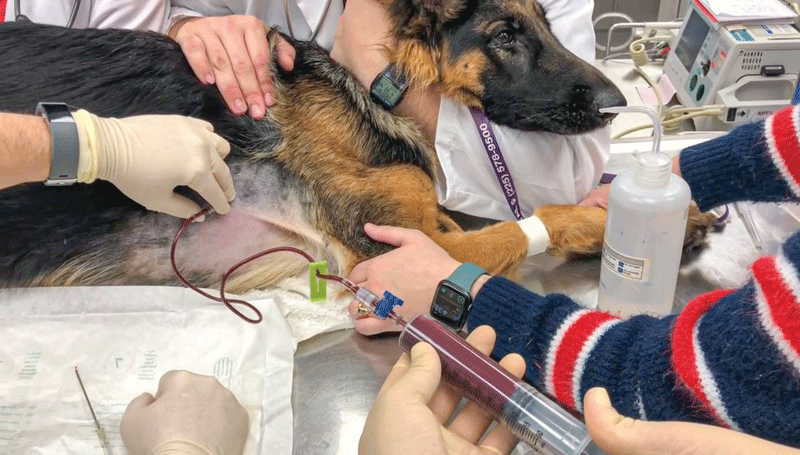
- In the emergency room. When performing an autotransfusion in the emergency room, the centesis site should first be clipped and surgically prepped. Both pleural and peritoneal cavities are appropriate sites for collecting autologous blood. The material needed to collect the blood is usually available in most veterinary practices and includes needle or over-the-needle catheter, three-way stopcock, extension line, and 60-cc syringes (Figure 1).
If a sterile collection bag is available, it can also be filled directly by using a three-way stopcock and two extension lines. Using sterile techniques, a needle or catheter can be inserted into the pleural or peritoneal cavity and blood collected directly into 60-cc syringes using the three-way stopcock and the extension line. The 60-cc syringes filled with autologous blood can be used for immediate blood administration into a peripheral vein of the patient using a blood filter or can be stored caped in the fridge for up to 24 hours.
- In the operating room. In some circumstances, blood can be collected directly during surgery. Blood cell salvage devices exist and can be utilized to collect the blood, wash it, and return it to the patient.
However, these devices remain very rare in veterinary medicine and a two-syringe technique, described by Robinson, presents a great and simple alternative method.1 In this method, a 60-cc catheter tip syringe is used to retrieve the blood from the peritoneal cavity. A luer tip 60-cc syringe is then connected to the catheter tip syringe and the blood transferred from the collecting syringe into the second (Figures 2 and 3). The 60-cc luer syringes can then once again be used to transfuse the patient immediately using a blood filter or can be stored in the fridge for up to 24 hours.
Possible complications
The use of autologous blood is associated with less transfusion reaction than allogenic blood products, however, hemolysis, coagulation disorders, micro embolism of fat or air, microaggregate of platelets, and sepsis are all complications that have been described following autotransfusion. In a 2015 retrospective study reviewing 25 cases of canine autotransfusions, 26 percent of the dogs showed hemolyzed serum post transfusion.2 This finding was, however, not associated with any clinical evidence of kidney dysfunction or other complications.
Autotransfusion represents a simple, cheap, safe, and life-saving alternative to the use of stored blood products in our companion animals and can easily be performed in an emergency setting.
Virginie Wurlod, Dr. vet. Med. DACVECC, DECVECC, is an associate professor in small animal emergency and critical care (ECC) at the Louisiana State University. Dr. Wurlod is originally from Switzerland, where she did her initial veterinary training, before pursuing an ECC residency in the U.S.
References
- Robinson DA, Kiefer K, Bassett R, et al. Autotransfusion in dogs using a 2-syringe technique. J Vet Emerg Crit Care (San Antonio). 2016 Nov:26(6):766-774.
- Higgs VA, Rudolff E, Kirby R, et al. Autologous blood transfusion in dogs with thoracic or abdominal hemorrhage : 25 cases (2007-2012). J Vet Emerg Crit Care (San Antonio). 2015 Nov-Dec;25(6):731-8.
- Kisielewicz C, Self IA. Canine and feline blood transfusions: controversies and recent advances in administration practices. Vet Anaesth Analg. 2014 May:41(3):233-42.