Radius and ulna fractures are common in small breed dogs, where they frequently affect the distal diaphysis. Traumatic radius and ulna fractures often affect the mid diaphysis in dogs and cats, but can occur at any location. Due to the limited soft tissue coverage of the middle to distal radius, open traumatic fractures can occur.
The initial consultation
When a patient first presents with diaphyseal fractures of both the radius and ulna, there will be gross instability; and due to the minimal soft tissue coverage, crepitus is often readily palpable. To decide the recommended treatment for the patient, it is important to accurately determine the signalment and history.
Age: A skeletally immature patient will have excellent healing capacity, with most dogs under six months old healing in six to 12 weeks, when sufficient stability is provided. However, if there is malalignment, malunion, and any damage to the physes, then as the patient continues to grow, this will lead to developing an angular limb deformity. It can be mind-blowing how quickly kittens and puppies less than 10 weeks old can heal. A bony callus can develop within 10 days and, therefore, if surgical intervention is planned, it should not be delayed.
A skeletally mature patient will take between two to five months to heal, and this will impact the decision on how to rigidly stabilize the fracture since external fixation or external coaptation for five months will require many rechecks, sedatives, bandage changes, frame cleanings, etc.
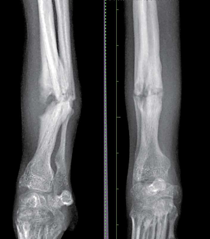
Breed: Toy breed dogs are predisposed to suffering distal radius and ulna fractures.1 Unfortunately, toy breeds have a high rate of radius and ulna fracture nonunion (80 percent or more), particularly if the fractures are treated with external coaptation (Figure 1).2 This high rate of nonunion is multifactorial, but is primarily thought to be due to reduced vascular density and reduced vessel arborization of the distal radius in toy breed dogs.3 This means that without rigid fracture fixation (typically internal fixation with a plate and screws), these fractures will not form a bony union.2
Species: It is paramount to remember cats are not small dogs, and their anatomy and function differ considerably such that specific considerations must be given to cats in cases of radius and ulna fracture.
One of the most noticeable differences between cats and dogs is cats perform a lot more supination of their paw, which requires mobility between the radius and ulna. This mobility of the bones relative to each other means traumatic fractures in cats can affect the ulna or radius in isolation more commonly than seen in dogs. It also means the radius and ulna are not able to internally splint each other as they can in the dog; therefore, each individual bone requires rigid fixation when fractured.
Cats can suffer from patella fracture and dental anomaly syndrome (PADS), which can lead to non-traumatic fractures of the olecranon.4 Therefore, assessment for concurrent non-traumatic fractures and persistent deciduous teeth should be performed.
Making a treatment plan
It is easy to focus on the fact the leg is dangling there and something should be done to fix it right away. While surgery may seem like the logical next step, it is wise to stop and consider whether any other injuries could be present that need treatment before definitive treatment of the fracture is performed.
Concurrent injuries: Traumatic radius and ulna fractures caused by blunt force trauma, such as being hit by a car or those sustained from a fall from a height can often be associated with concurrent injuries. Patients should be assessed for the presence of thoracic, abdominal, and neurological injuries.
A complete and thorough clinical examination can aid in assessing whether there are rib fractures, possible pneumothorax, abdominal wall hernias, and if the patient is in shock.
A full neurological examination should be performed, with a focus on ruling in or out head injury, neck fractures, and brachial plexus injury.
Horner’s syndrome, loss of cutaneous trunci or a miotic pupil can indicate brachial plexus injury and further investigation into this should be performed if present. The radial nerve is essential for thoracic limb weight bearing in the dog due to its innervation of the triceps and extensor carpi radialis muscles.5
In many dogs with antebrachial fractures, it is very difficult to perform a withdrawal and spinal reflexes due to the pain they are in. Thus, assessing whether the radial nerve is severely damaged can be done by assessing superficial sensation to the dorsal surface of the paw, which is innervated by the radial nerve.
While covering the eyes of the patient, take a pair of small hemostats and gently pinch the skin on the dorsal paw of the affected leg. If positive, the patient will actively try to move the leg away from you and/or turn towards you to signal they want you to stop pinching their paw. This indicates superficial sensation of the dorsal paw is present and in most cases with this sensation, full radial nerve function will be recovered within six weeks.
Lack of superficial sensation should prompt detailed neurological examination to determine the severity of injury and the prognosis for return to function. Although be aware some dogs in shock after the trauma or following large amounts of opioids may have a false negative on this test.
Thoracic and abdominal radiographs and ultrasound plus an electrocardiogram will allow assessment for rib fractures, pneumothorax and hemothorax, diaphragmatic hernia, myocardial damage (arrhythmias), abdominal wall hernias, urinary tract rupture, and abdominal hemorrhage.
A full orthopedic examination can help assess presence of any concurrent fractures; although in the case of atraumatic fractures in cats, survey radiographs may be required due to a lack of associated soft tissue swelling making them more difficult to detect. Falls from a height can frequently be associated with ligamentous injury, so particular attention should be paid to assessing the ligamentous support of the carpi and tarsi of these patients.
During your investigation of concurrent injuries, you may want to place a temporary soft-padded bandage +/- a splint on the fractured antebrachium to aid patient comfort, prevent any further contamination if wounds are present.
Radiographs of the fracture: Craniocaudal and mediolateral views, including the elbow and carpus, are usually sufficient for diagnosis of most radius and ulna fractures. In highly comminuted fractures, oblique views or a computed tomography scan may be useful to further assess for the presence of fissures that may affect the treatment plan, particularly if the fissures extend close to the antebrachiocarpal joint. It is usually easier to attain straight views without a bandage in place, however, if a wound is present then this should be covered with a dressing and light bandage to prevent further contamination.
Splint, cast, or surgery?
The decision on a definitive treatment plan should consider the biology of the fracture, the mechanics of the fracture, and various clinical factors, including patient temperament and client budget. A detailed description of the treatment options and their risks versus benefits is beyond the scope of this article, but three key points are below:
1) Any toy breed dog with a radius and ulna fracture should undergo surgical fixation, usually rigid internal fixation, and should not be treated with a cast or a splint. With the increasing popularity of toy poodle-mixed breeds, this should be extrapolated to any breed that has a “toy breed-like” antebrachium, such as mini labradoodles, Pomskies, Italian greyhound crosses, etc. Consideration of the size of the implant used for these dogs is highly important as there is a risk of osteopenia occurring with an oversized implant.6
2) Young animals are at risk of developing an angular limb deformity after suffering a radius and ulna fracture. This can be worsened if overlong screws are used in the proximal radius and a synostosis is created.
Similarly, a synostosis between the radius and ulna can occur if there is too much instability and a large callus occurs, most commonly seen with external coaptation.
Although young animals can heal quickly if both bones are fractured, it may be best to advise surgical fixation to minimize the risk of synostosis (remembering to keep those proximal screws short) rather than to splint or cast the leg. However, if only the radius or only the ulna is fractured in a young patient, then the chance of synostosis is considerably reduced and external coaptation can be very successful.
3) Cats do not tolerate thoracic limb splints or casts well (they tend to “shake” them off before they even make it home). There is also a lot of mobility between the radius and ulna, which is important to preserve, but reduces the overall stability of antebrachial fractures even if only one of the bones is affected. Therefore, most feline antebrachial fractures benefit from surgical fixation.
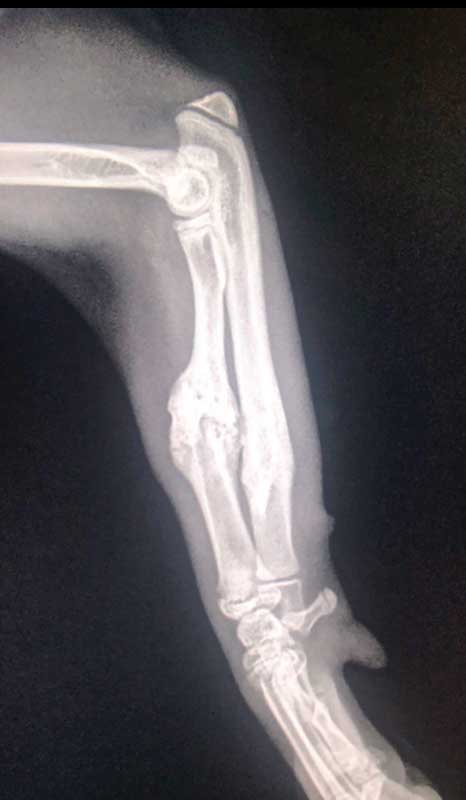
However, very young, healthy kittens have phenomenal healing capacity; I had a six-week-old kitten that we tried to splint as surgery was not an option due to some other fractures that we had to prioritize; and after only two weeks, the fracture had completely healed despite him only tolerating his splint for one day! (Figure 2)
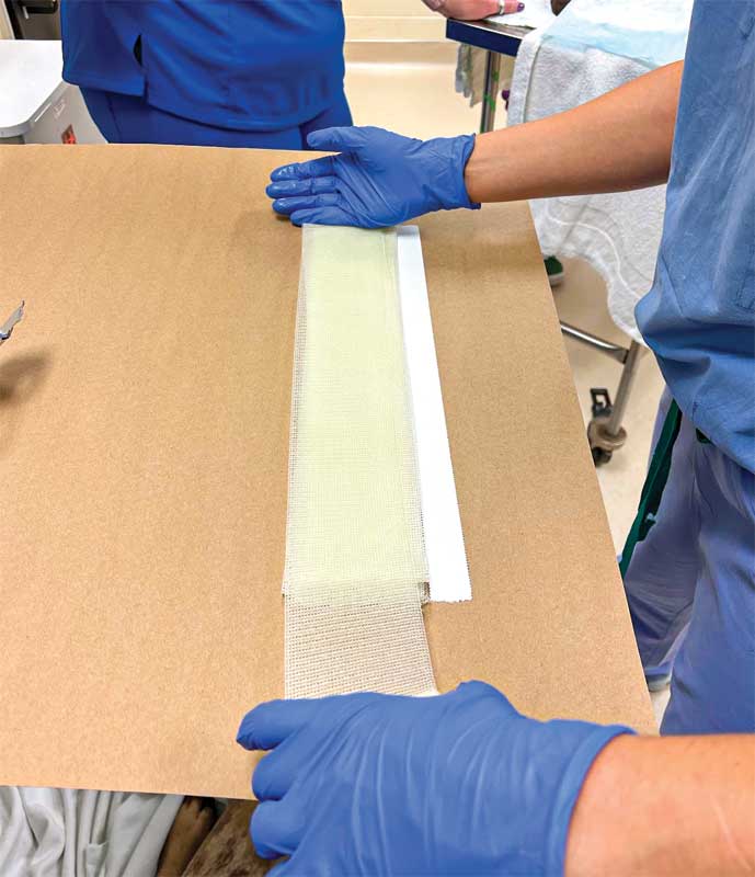
Bonus point: Young, healthy dogs with only the radius or only the ulna fractured, with minimal displacement, closed fractures can do great with six to 12 weeks of splinting. We tend to use cast tape (Figure 3) to customize a lateral splint for each case so we can stabilize the elbow (you need to stabilize the joint above and below for effective splinting/casting). Using the cast tape allows us to avoid having to place a full cast since we can make a strong, custom-fitting splint for each patient that we can adjust as required using a cast saw to round off edges, etc.
Jessica McCarthy, BVSc, MRCVS, DECVS, works as a faculty in orthopedic surgery at the University of Wisconsin. Dr. McCarthy became a diplomat of the European College of Veterinary Surgeons in 2020, after completing her residency training at the University of Edinburgh. McCarthy attended Bristol University for her veterinary degree and spent two years in general practice in England after graduating. She has a particular interest in elbow disease, both developmental and traumatic, and is passionate about ensuring everyone in the veterinary profession can work in a diverse and inclusive atmosphere.
References
- Harasen G. External coaptation of distal radius and ulna fractures. Can Vet J. 2003 Dec;44(12):1010–1.
- Sumner-Smith G, Cawley AJ. Nonunion of fractures in the dog. J Small Anim Pract. 1970 May;11(5):311–25.
- Welch JA, Boudrieau RJ, DeJardin LM, Spodnick GJ. The intraosseous blood supply of the canine radius: implications for healing of distal fractures in small dogs. Vet Surg. 1997 Feb;26(1):57–61.
- Reyes NA, Longley M, Bailey S, Langley-Hobbs SJ. Incidence and types of preceding and subsequent fractures in cats with patellar fracture and dental anomaly syndrome. J Feline Med Surg. 2019 Aug;21(8):750–64.
- Hermanson JW, Evans HE, de Lahunta A. Miller and Evans’ anatomy of the dog-E-book. Elsevier Health Sciences; 2018.
- Larsen LJ, Roush JK, McLaughlin RM. Bone plate fixation of distal radius and ulna fractures in small- and miniature-breed dogs. J Am Anim Hosp Assoc. 1999 Jun;35(3):243–50.