By Anne S. Hale, DVM • VetStem, Inc.
Today, the veterinary practitioner has many product choices for regenerative medicine. It is important to understand how and why these products work in the area of interest to maximize your patient’s opportunity for success. Biologics, including cell-based products, are available both as an autologous and allogeneic formulation. A review of how cells, specifically platelets, work to promote injury repair and inflammation resolution is necessary to support the correct use of platelet-rich plasma.
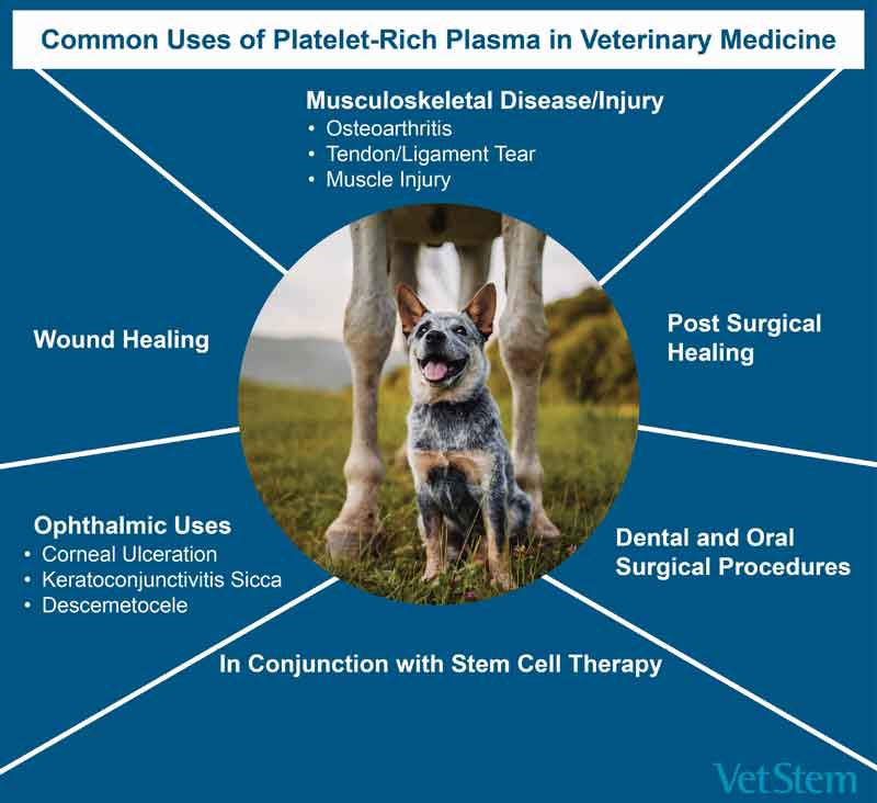
What is PRP and How Can it be Used?
By definition, platelet-rich plasma (PRP) is a concentrated platelet product with native plasma. The purpose for platelet concentration is to provide a higher number of platelets than you would find in normal blood circulation in an effort to enhance local angiogenesis, regulate cellular activity, home stem cells, proliferate stem cells, and deposit matrix proteins to support healing and regeneration of tissue.1
Published uses of PRP in the dog and horse include repair and regeneration of muscle, ligament, tendon, bone, dermis, epidermis, and cornea. Most commonly, veterinary clinicians have experience with PRP and its uses for musculoskeletal diseases and injuries.2 Its use in large wounds and burns has also been well documented. Corneal ulceration, keratoconjunctivitis sicca, and descemetocele are frequent ophthalmic uses of PRP. Historically, surgeons have placed PRP at the site of fracture repair to enhance healing post-surgery. More recently, PRP has been utilized intra-cavitary to address inflammation, for example endometritis or chronic cystitis. The veterinary practitioner has long since seen these treatment modalities as a way to “home” stem cells.
Recognizing the broader role that the platelet plays in endothelial stabilization and hemostasis, PRP has also been explored in laminitis, sepsis, hemorrhagic shock, and traumatic brain injuries. Each of these disease indications rely on the platelet to support an anti-inflammatory environment while participating locally in the stabilization of the local hemostatic environment.
Largely through topical or intra-articular applications, PRP has been used to provide local analgesia and sourcing for growth factors. In addition, these applications have supported an antibacterial role for the PRP through its ability to signal other cell types.
Factors Supporting Successful PRP Therapy
Successful PRP therapy is based on total platelet dose, the functionality of the platelet membrane, and ability to degranulate. Published reviews and clinical studies utilizing autologous PRP produced by a medical device show that variability in platelet number, platelet activity, and platelet phenotype lead to variable clinical outcomes.
Allogeneic pooled platelet-rich plasma offers an opportunity to overcome these concerns by providing a consistent platelet number, minimizing premature activation and degranulation, as well as controlling donor variability. Once this variability has been overcome, then successful PRP therapeutics are based on an overconcentration of platelets in the lesion, joint, or topical location. A review of PRP therapy and clinical outcomes written by Carmago Garbin et al. supports that five times the normal circulating platelet count is ideal for positive outcomes.3
PRP Dosing Considerations
Dosing PRP requires an understanding of the disease indication as well as the location. For instance, if the area to be treated is a synovial membrane and the subintimal connective tissue layer, then PRP should be dosed for maximum effect to allow for “bathing” of the cavity and penetration into the subintimal layer. Intra-articular administration allows for delivery to the lesion without dependency on intravascular circulation as well as local concentration of product. Published human reports on intra-articular use of PRP suggests that approximately 2.5 billion platelets are needed to provide the appropriate total platelet dose for positive outcomes.4
When treating intralesional defects, dosing is affected by the size of the lesion as well as its location and time from injury. These lesions, such as superficial digital flexor tendon tears or supraspinatus tendinopathies, may be treated with a similar total platelet dose. Published randomized trials in equine patients with superficial digital flexor tendinopathies support a total platelet dose of 2.7 billion platelets divided into three sites with positive outcomes.5
Topical dosing must be calculated depending on the lesion size, containment in the lesion, and activation level of the platelets. This type of use is focused on exposing the lesion to high quantities of growth factors to promote regeneration and angiogenesis. Strategies for maintaining contact in these areas include adding fibrin, synthetic gels, and/or topical creams to “hold” the platelets in the area until degranulation. Specifically, corneal ulceration therapeutics involve subconjunctival injections of PRP once weekly for two to three treatments. In a study of 28 dogs/cats, subconjunctival injections were successful in promoting resolution in corneal ulceration when given two to three times at a frequency of once weekly.6
Conclusion
In studies where the platelet count can be controlled and site administration is localized, positive outcomes with the use of PRP can be expected. Allogeneic PRP has the advantage of being an off-the-shelf therapeutic with a known and consistent platelet dose and potency. This provides an easy and convenient way to administer PRP therapy to your patients for a variety of disease indications.
References
1 Harrison P, Subcommittee on Platelet P. The use of platelets in regenerative medicine and proposal for a new classification system: guidance from the SSC of the ISTH. J Thromb Haemost. Sep 2018;16(9):1895-1900. doi:10.1111/jth.14223
2 Sharun K, Jambagi K, Dhama K, Kumar R, Pawde AM, Amarpal. Therapeutic Potential of Platelet-Rich Plasma in Canine Medicine. Arch Razi Inst. Oct 2021;76(4):721-730. doi:10.22092/ari.2021.355953.1749
3 Camargo Garbin L, Lopez C, Carmona JU. A critical overview of the use of platelet-rich plasma in equine medicine over the last decade. Frontiers in veterinary science. 2021;8:641818.
4 Raeissadat SA, Ghazi Hosseini P, Bahrami MH, et al. The comparison effects of intra-articular injection of Platelet Rich Plasma (PRP), Plasma Rich in Growth Factor (PRGF), Hyaluronic Acid (HA), and ozone in knee osteoarthritis; a one year randomized clinical trial. BMC musculoskeletal disorders. Feb 3 2021;22(1):134. doi:10.1186/s12891-021-04017-x
5 Geburek F, Gaus M, van Schie HT, Rohn K, Stadler PM. Effect of intralesional platelet-rich plasma (PRP) treatment on clinical and ultrasonographic parameters in equine naturally occurring superficial digital flexor tendinopathies – a randomized prospective controlled clinical trial. BMC Vet Res. Sep 7 2016;12(1):191. doi:10.1186/s12917-016-0826-1
6 Farghali HA, AbdElKader NA, AbuBakr HO, et al. Corneal Ulcer in Dogs and Cats: Novel Clinical Application of Regenerative Therapy Using Subconjunctival Injection of Autologous Platelet-Rich Plasma. Frontiers in veterinary science. 2021;8:641265. doi:10.3389/fvets.2021.641265