Many veterinary professionals are frustrated with dentistry, and some of that frustration stems from not having the correct equipment, improper instrument maintenance, and a lack of training. Having the right instruments, and keeping them well-maintained, can make a huge difference.
Periodontal instruments
The most common procedure in veterinary dentistry is a complete dental cleaning and polishing, with full mouth examination, including intraoral radiographs. While most of the dental cleaning is done with mechanical scalers (discussed in “Tips to build your dental suite operatory”), the proper periodontal hand instruments are typically underutilized.
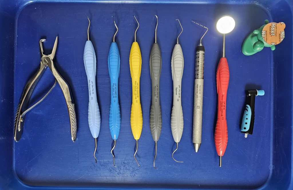
It is important to have good periodontal packs, sufficient for the number of procedures expected to be done in a day (Figure 1). These periodontal packs should include:
1) Periodontal probe and explorer. Each is often on opposite end of a double-ended instrument (Figure 2).
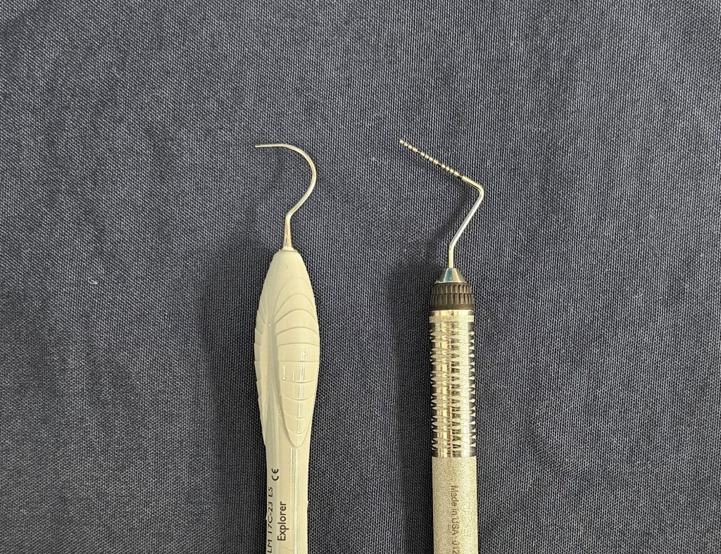
- The probe is used to measure pocket depth around the tooth. It measures any extent of root exposure caused by gingival recession. Total attachment loss is determined by adding the root exposure in millimeters to the pocket depth in millimeters, or the distance from the neck of the tooth to the depth of the periodontal pocket.The probe may also measure any “pseudopocket” formed with gingival enlargement or hyperplasia. It can be used to determine if there is any furcation exposure (mild, halfway through, or all the way through) with attachment loss at multi-rooted teeth. Note: There are many variations of markings on probes; be sure to know what your probe measures in millimeter increments (Figure 3).
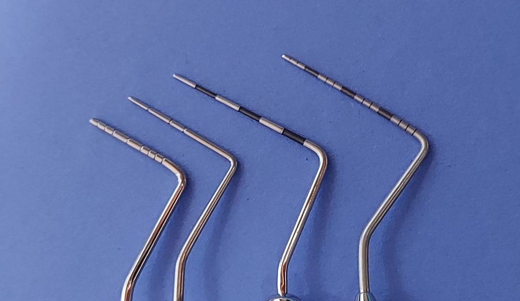
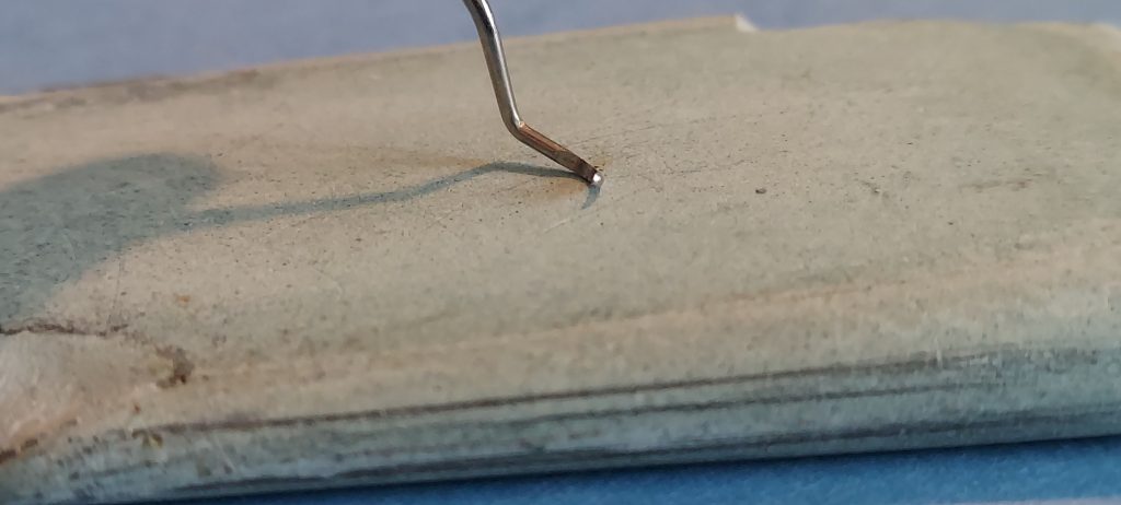
Figure 3.There are many types of periodontal probes, often with different marks for measurements in millimeters. - The sharp tip of the explorer (often shaped like a shepherd’s hook) is a tactile instrument to feel for any remaining calculus or irregularities in the enamel, such as tooth resorption, carious lesion or pulp exposure due to a fractured tooth.
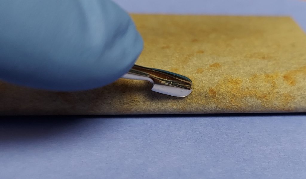
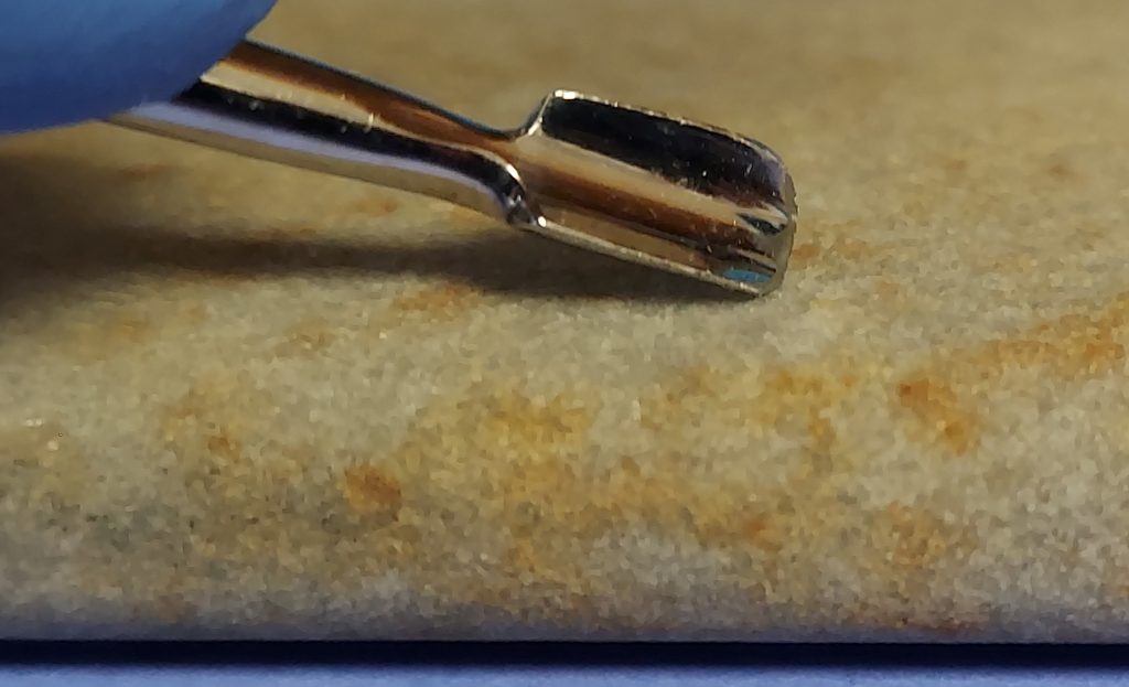
2) Hand curettes are probably the most under-utilized dental instrument. With its rounded toe and rounded back, it can be inserted into the gingival sulcus or pocket to scale the root surface hidden under the gumline.
Most curettes have a slight angle of the working head, often with a mirror image on the opposite side of the instrument. In contrast with the angled blades of Gracey and McCall curettes (among others), a universal curette’s face is 90 degrees to the handle. With the face of the instrument “closed” (facing the tooth), this allows one edge of the curette to contact the root surface; as it is pulled out, it scrapes the calculus and debris from the root surface. (Figure 4).
Each end can be used for various surfaces of the teeth, and the different shapes allow for better contact on various tooth surfaces.
3) Hand scalers have a sharp tip and are often triangular in cross-section. Because of their sharp edges, they should not be used subgingivally, but the tip is useful in cleaning grooves. The hand scaler can be used on any supragingival surface.
4) Dental mirrors can be very useful in visualizing the distal surfaces of molars during examination and cleaning. However, they are also underutilized.
5) Calculus forceps can help in the careful removal of gross calculus from the crown surfaces, but you must take care not to damage the tooth.
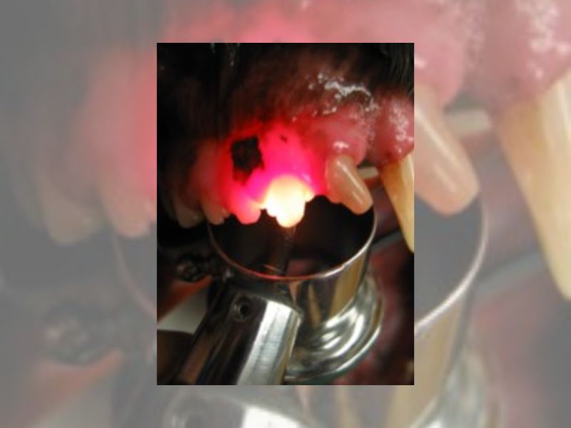
6) Having a bright penlight as a transillumination can be a useful part of your full dental examination. With any chipped, broken or discolored tooth, shining the light from behind the tooth may help you visualize changes that indicate a non-vital pulp (dark, does not allow light through the tooth). Compare to similar teeth; if the tooth is vital, it should allow the light to shine through and even show the pink of the vital pulp. (Figure 5)
Extraction instruments
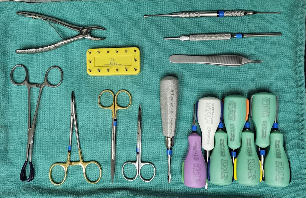
In addition to the highspeed dental units, handpieces and burs, oral surgery sets of hand instruments can be put together, organized and color-coded to provide options for cats and small dogs to large dogs and oral surgery soft tissue sets (Figure 6).
General surgery items are used, such as scalpel blade handles, scalpels (15 or 15C), forceps (small, delicate ones for soft tissue handling), scissors (suture scissors and sharp scissors for tissue) and needle drivers (personal preference, but usually smaller instruments). In addition to these, specific instruments for extraction and oral surgery are helpful.
- Periosteal elevators are necessary to help elevate gingival flaps and to debride alveolar sockets and soft tissue (Figure 7). Serrated periosteal elevators are useful for debridement as well. (Figure 8).
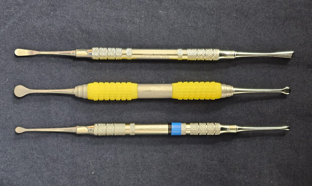
Figure 7. From top: EX7 (similar to a Howard periosteal elevator), EXPM, and Molt #2/#4. 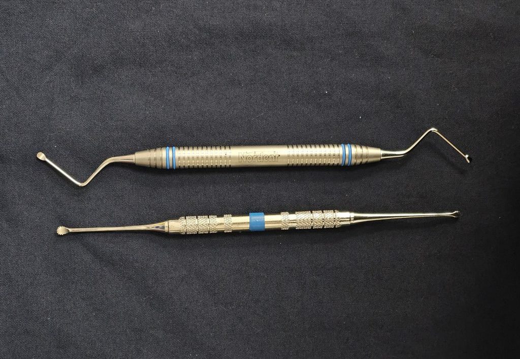
Figure 8.A serrated periosteal elevators and bone curette for debridement. - Dental elevators and luxators are blade‐tipped instruments that lever the tooth out of its socket. They are designed to be used in the periodontal ligament space for maximal stretching of the periodontal ligament (PDL), so a sharpened tip is essential for placement in the space.Luxator tips are generally flat and more delicate than elevators and are used initially to severe the PDL attachment, so there should be no rotation of the instrument (Figure 9).
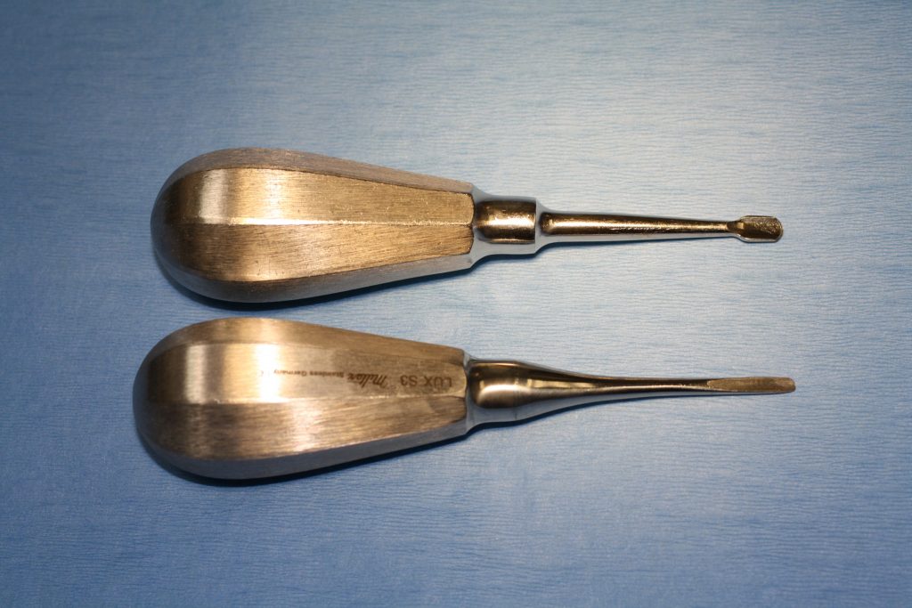
Figure 9.The difference between a flat, thin luxator, and winged elevator. Some larger and thicker luxators can be useful on the flat surfaces of large teeth such as canines. Elevators are thicker and can withstand appropriate forces needed for leverage. There are different sizes of winged elevator to correspond to fit the size and diameter of the root (Figure 10).
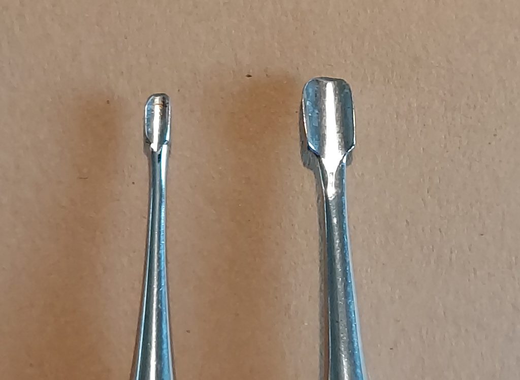
Figure 10. Winged elevators come in different sizes to correspond to the size of the root to be extracted. Different handle sizes are also available to help with operator grip. Smaller elevators and luxators and even some root tip picks are designed for small teeth and roots (Figure 11).
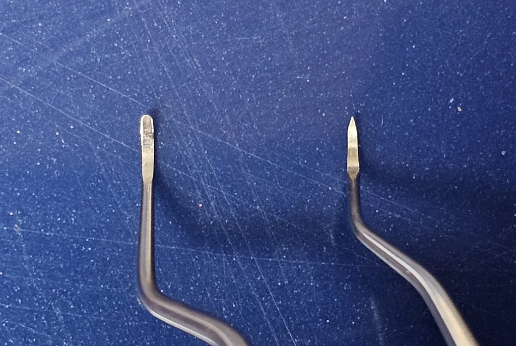
Figure 11.Smaller elevators, luxators, and root tip picks can be used to help retrieve and elevate fractured roots. - Small breed extraction forceps are preferred, to avoid excessive forces when removing the loosened tooth/tooth segment. They can be gently used to rotate the segment to ascertain the need for any continued elevation or bone removal (Figure 12).
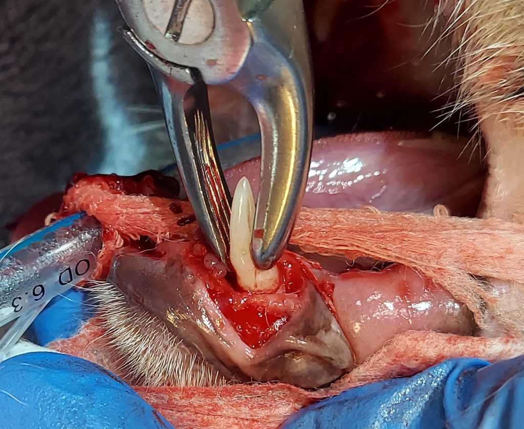
Figure 12.Small breed extraction forceps can be used to assess the extent of PDL fatigue as well as grasping the crown for full elevation. - Lip retractors can be very useful to hold back lips for access to the surgical field. Even spiral perm rollers can be used as soft mouth gags, to hold the lips back and even hold radiograph sensors in place (Figure 13).
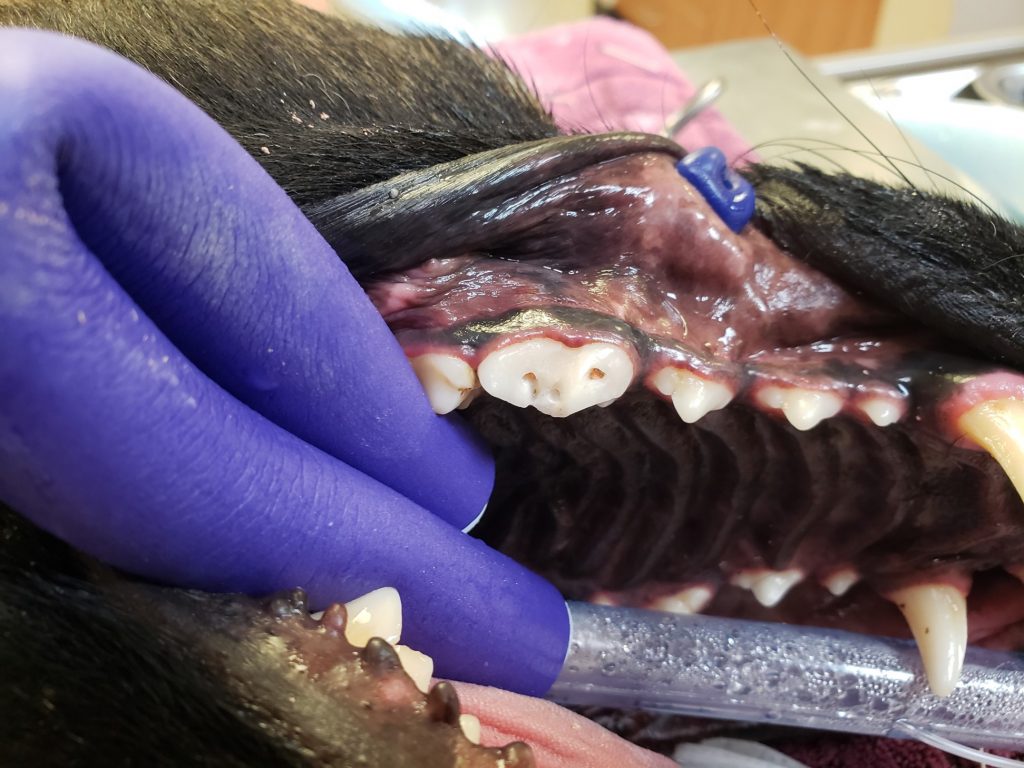
Figure 13.During this root canal procedure, a spiral perm roller was folded and used both for a soft mouth gag and to help retract the lips, and the tip of a lip retractor can be seen holding back the upper lip. - Suture material should be absorbable and monofilament, as braided material can wick bacteria into the site. While some use chromic catgut, these authors prefer polyglycolide-co-caprolactone, 4/0 with reverse cutting needle for dogs, and 5/0 with tapered needle for cats.
Additional equipment may include a directed power-cutting instrument such as a piezotome that provides variable-intensity piezoelectric vibrations. Used with an up-and-down motion in the PDL space, this can help reduce strain on the operator, preserving soft tissue with minimal bone loss as compared to needing osseous resection. Advanced units are also used in oral surgery with osteotomy procedures.
Instrument sharpening
A dull instrument, whether it is a hand curette, periosteal elevator, or dental elevator, is not an efficient tool. With the curette, a dull instrument can just burnish the calculus instead of removing it, and a dull dental elevator with its thick, dull blade will not be effective for PDL space placement. A simple flat Arkansas stone, or one with round edges or grooves, may be all you need to keep your instruments in optimal shape. A Bates Arkansas stone has several grooves placed in the surface to help in sharpening winged elevators. Honing machines are fast, but excessive force can damage the instruments, so care is needed.
Hand curettes typically only need one edge on each working head to be sharpened, the edge that contacts the tooth surface. There are several techniques, including the moving stone to the moving instrument technique,1,2 each positioning the blade edge at a 110-degree angle to the stone (Figure 14). The curette is sharpened from the heel to the toe of the instrument, taking care to keep the toe rounded. Scalers should have sharp edges and even the sharp tip maintained.
Dental elevators need to have the tip of the instrument with a blade sharp enough to enter the PDL space. Several techniques are also possible with these instruments, including sharpening the face of the instrument on a rounded edge of a stone, holding it at a 30- to 45-degree angle (Figure 15). These can also be sharpened on the back side on the flat surface of a stone, or within the grooves of a Bates stone made for winged elevator sharpening (Figure 16).
Periosteal elevators generally do not need as much sharpening but should be examined regularly for any defects or dents in the metal surfaces, to keep a sharp edge.
The most important aspect of sharpening is that each instrument should be examined after each use, and if the edge is dull, the instrument should be sharpened. Waiting for the “sharpening service” once a month is not adequate. In addition, with each sharpening, some of the metal of the instrument is removed. With dental elevators, this means that the gradual loss of the thinner tip will make the instrument head shorter, and the metal is generally thicker towards the handle of the instrument. Once this happens, a sharp edge may be produced on the tip, but the general thickness will not allow easy insertion into the PDL space.
As curettes are sharpened, the working head can become quite thin and fragile, and can even break with use. So as much as veterinary teams like to hang on to equipment for as long as possible, it is essential to replace old, worn-out or damaged instruments on a regular basis. For a select few pieces, they may be used as tissue retractors or protectors, what we affectionately call “zombies,” using the old instrument to keep tissues from being damaged from highspeed burs (Tip: Do not use your good instrument for that.).
In summary, without the right instruments that are properly cared for, periodontal therapy, extractions, and oral surgery will be challenging at best and frustrating for many. Once you have the right tools, you may wonder how you ever managed before!
Heidi Lobprise, DVM, DAVDC, is a 1983 Texas A&M University graduate. She became board-certified in dentistry in 1993. After 10 years in the industry, Dr. Lobprise returned to dental specialty practice in 2014 and has since “semi-retired” in Kerrville, Tex. Lobprise is the author/co-author of three dental texts, writing many chapters and articles.
Shannon VanTrease has been in the veterinary field for more than 25 years. He has a wide variety of experience, including emergency medicine, companion animal practice, large animal medicine, and has spent around 15 years in veterinary dentistry. VanTrease’s interest in veterinary dentistry began during his time working for Robert Wiggs, DVM, DAVDC, followed by Heidi Lobprise, DVM, DAVDC, and now the current dental team at the Flower Mound Veterinary Emergency & Specialty Center in Flower Mound, Tex. This has given him the opportunity to work with four generations of veterinary dentists. VanTrease enjoys educating technicians, students, residents, and veterinarians. He is looking forward to providing education to a larger audience with the addition of a new veterinary dentistry training facility opening in 2025.
References
- Hu-Friedy’s It’s About Time | Sharpening Gracey Curettes (youtube.com). https://www.youtube.com/watch?v=Y4s_q_AdK38
- Gracey Curette Sharpening Stationary Stone & Moving Instrument (youtube.com). https://www.youtube.com/watch?v=l2OgnR_VLKY&t=3s (@nancyFoster870)