Ultrasound is a mainstay diagnostic tool for many practitioners, but for Nathan C. Nelson DVM, MS, DACVR, DACVR-Equine Diagnostic Imaging, ultrasound is also the tool that often gets his team answers they could have not had with other modalities.
“Our most common use is abdominal imaging in patients with diseases that are not apparent on abdominal radiographs or when radiographs do not give us a definitive answer,” says Dr. Nelson, clinical professor, diagnostic imaging, Department of Molecular Biomedical Sciences, College of Veterinary Medicine, North Carolina State University.
Nelson also often uses ultrasound to guide organ sampling for cytology analysis and culture, such as when the practitioners identify liver nodules, sampling some of the nodules using a needle guided by ultrasound.
Lastly, Nelson finds ultrasound to be “ideal for evaluating function,” such as echocardiographs to evaluate for cardiac function, or to evaluate blood flow within a structure (i.e. looking for a thrombus within a vessel).
Ultrasounds can also provide real-time evaluation of blood vessels and peristalsis, which provides “a large amount of clinical information” to help practitioners better understand their patient’s problems and better care for them, according to Tony Pease, DVM, MS, DACVR, chief veterinary medical officer of Viticus Group – Animal Health, a tenured associate professor and section chief, Veterinary Diagnostic Imaging at Michigan State University, and president of the American College of Veterinary Radiology (ACVR).
“I personally really like using ultrasound to evaluate intestinal foreign bodies,” says Dr. Pease. “You can evaluate the small intestinal wall, look for evidence of perforation and assess partial obstructions. Ultrasound is also great at looking for pancreatitis, although it takes some training. Ultimately, the best use of ultrasound is to guide needles to get aspirates of lesions so we can understand what the lesion is and help with treatment and prognosis.”
When Grant Middleton, DVM, DACVR, with Vision Pet Imaging, a diagnostic imaging group in the greater Puget Sound region, performs a complete abdominal scan, he and his team are looking at the liver, the spleen, the kidneys, the bladder, reproductive organs, adrenals, the upper and lower urinary tract, the pancreas, the GI tract, lymph nodes and vessels in the abdomen.
“So, primarily we’re using ultrasound for further assessment of intra-abdominal pathology,” says Dr. Middleton, who is also president of the Veterinary Ultrasound Society (ACVR).
Less common uses he lists are: Checking for cervical disease by examining thyroids and parathyroids and salivary glands; cervical lymph nodes; musculoskeletal disease, specifically the shoulder (or) calcaneal tendons; ocular scans; as well as the diagnostic tool used by cardiologists anytime they perform an echocardiogram.
“Frequently dogs will come into the veterinary clinic with pretty nondescript clinical signs and blood work and imaging makeup, you know the majority of the diagnostic tools that we begin with trying to isolate what problems exist, so once blood work abnormalities occur, especially liver enzyme or kidney enzyme elevations or signs of infection or signs of anemia and blood loss,” Middleton adds. “Those types of things clue us into a problem that may potentially be occurring in the abdomen.”
The next diagnostic step is to differentiate whether the issues are related to infection or merely an inflammation, according to Middleton.
Radiographs can identify some foreign material, but there is a lot of foreign material that cannot be seen as well on an X-ray, so an ultrasound may help a practitioner understand if more definitively whether there is evidence of foreign material, an obstruction, or if it’s just primary pancreatitis, Dr. Middleton adds.
On most days, Anthony Fischetti, DVM, MS, DACVR, and his staff perform about 30 ultrasounds.
“It never ceases to be interesting,” says Dr. Fischetti, senior veterinarian, specialist in radiology, diagnostic imaging, service head of diagnostic imaging, Diagnostic Imaging Residency Program Director Schwarzman Animal Medical Center, in New York. “On any given day, we identify obstructive foreign bodies in pets’ GI tracts.”
Some cases lead them to problems that can be easily treated, and a good outcome for the patient. Other problems identified portend poorly for patients.
“We unfortunately identify cancerous lesions every day, he adds. “We can then use ultrasound to get a sample of the mass for a cytological diagnosis.”
One advantage animal medicine in the use of ultrasound over the field of human medicine is ultrasounds performed on smaller pets can yield much more to help in diagnoses and ensuing treatments.
Fischetti works with a technician who has been a human ultrasound technician for several years. “And she is amazed at how much information we get from the abdomen of our cats and small dogs,” Dr. Fischetti adds. “For example, the GI tract to a sonographer of people is generally considered just ‘in the way,’ but the GI tract is a very important part of the dog/cat comprehensive abdominal ultrasound.”
Trends
A trend toward efficiency that can be found in the development of ultrasound has been with the introduction of handheld and highly portable ultrasound units.
“All of the electronics are located within the small, wireless probe,” says Dr. Nelson of the newer units. “The images are displayed on a handheld tablet nearby. There are some limitations to that type of unit, but they have improved greatly and are very reasonably priced.”
Pease, who also believes the increased accessibility of ultrasound equipment, says practitioners should view a “relatively inexpensive tool,” yet as one requiring some advanced training with a learning curve.
“I think the only reason veterinarians do not have an ultrasound is because they do not know how to use it,” Pease says, adding many companies provide ultrasound training courses to allow clinicians to bring the technology to the practice.
Middleton sees “a potential paradigm shift” in the field, in which the demand for ultrasound examinations has risen to the point it is higher than the ability of boarded radiologist to perform those scans.
In human medicine, numerous programs have been developed to give non- boarded radiologists a higher level of training, as well as certifications to provide a certain level of competency with ultrasound to meet growing demand, he noted.
“In our field, the demand for ultrasound is just so unbelievably high,” Middleton says, citing the development of training and credentialing programs. “So many cases that walk into a vet clinic really would benefit from an abdominal ultrasound and we’re seeing that become more the standard of care in these workups, especially with clients who are looking to actually try and seek what the root problem is and are willing to perform you know more advanced therapy is you know ultrasound becomes a pretty critical piece of the functioning of.”
Buying equipment
Portability and accessibility are key factors in purchasing equipment. Nelson believes affordability is also worthy of consideration.
“In general, you get what you pay for,” Nelson says. “While there are a large number of newer portable ultrasound units, in general, when you buy a larger laptop or even larger upright form factor ultrasound, you will see an improvement in image quality.”
Additionally, larger machines often have an array of ultrasound transducers providing additional capabilities beyond those afforded by cheaper or handheld units.
“For instance, with very big patients or deep-chested patients, you may need a larger more powerful transducer, which are usually available on the larger, more expensive units,” he says. “You have to decide what capabilities you need/want for your practice, and then choose an ultrasound machine that meets those needs.”
Nelson advises deciding what ultrasound will be used and choose one based on that. Will it be used primarily in a cat-only practice? “In that case, you really just need a high-resolution linear transducer.”
On the other hand, for practices performing equine orthopedic imaging, the types of probes you need for best imaging such areas will be different and will not be supported by all machines. “There are lots of vendors out there and you are not just buying a machine, you are becoming part of a company,” Pease says, advising considering trying multiple units. “Do you like their customer service? Do they have support? Do they have a tele-radiology company you can use that may help you get the most out of your machine?”
Don Jergler has been a reporter for more than 25 years, covering insurance, real estate, and other topics. He spent two decades as a reporter at several daily newspapers, then entered business-to-business reporting. Jergler is currently the Western Region editor of Insurance Journal and editor of
Claims Journal.
References
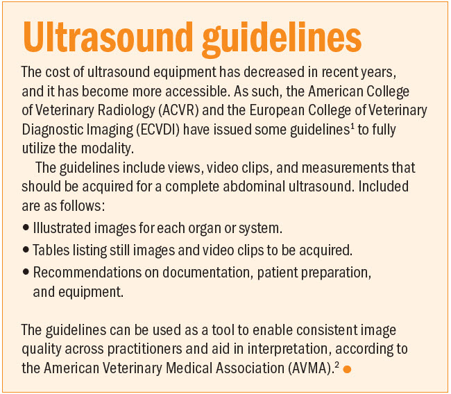
- https://onlinelibrary.wiley.com/doi/10.1111/vru.13151
- https://www.avma.org/news/specialists-issue-consensus-statement-abdominal-ultrasound-dogs-cats