Patients presenting to an emergency service with polytrauma can be very challenging. While severe visible injuries, such as degloving wounds, can cause distress to practitioners, threatening internal injuries are easily overlooked, leading to more compromising conditions, such as cardiopulmonary arrest.
Trauma types
Traumatic injuries can result from of a variety of causes, such as road traffic accidents, bite wounds, high-rise falls, or gunshot wounds. These injuries are of two types: blunt traumatic injury or penetrating traumatic injury. Both injuries can affect all organ systems, so assessing the whole body is important.
Epidemiologic studies in dogs and cats1-4 have shown trauma patients are mostly young males. Geriatric patients account for only nine percent of the presenting traumas, and only six percent of trauma patients have pre-existing illness. With appropriate care, survival to discharge rate for the trauma patient is usually very good (85-88 percent).
The most common traumatic injuries according to various studies are:
- Thoracic injuries: range from 38-72 percent (pulmonary contusions, pneumothorax, hemothorax, and rib fractures)
- Abdominal injuries: 12-50 percent (hemoperitoneum [23-38 percent], urinary tract rupture [two-three percent], abdominal hernia [five percent])
- Head injuries 24-25 percent
- Orthopedic injuries: fractures and luxations (87 percent)
- Soft tissue injuries: abrasions (56 percent), lacerations (26 percent), subcutaneous emphysema (10 percent) and major degloving (eight percent).
Primary survey of the trauma patient
When a polytrauma patient presents to the emergency service, an initial step-by-step approach is recommended using ABCD: Airway, Breathing, Circulation and Disability.
Airway and breathing
Airway and breathing can be assessed simultaneously in trauma patients. Primary airway obstruction or disease is uncommon in trauma patients, but can be observed with significant injuries to the head and neck.
On the other hand, abnormal breathing pattern is seen in a majority of blunt trauma patients. Visual assessment of the airways and breathing pattern and chest auscultation are the most effective way to localize and diagnose these problems. Additionally, pulse oximetry and thoracic focused assessment with sonography for trauma (tFAST) scan can help assess oxygenation and intrathoracic injuries.
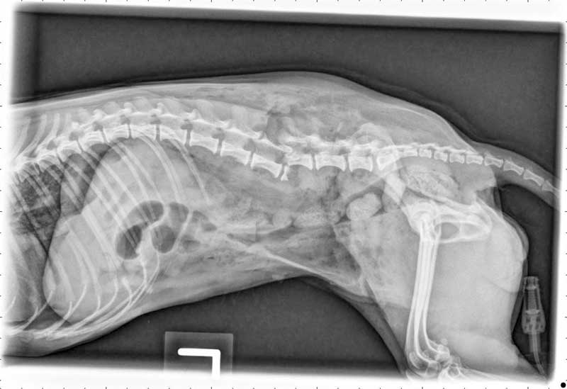
Thoracic radiographs should be performed in all polytrauma cases, but only if the patient is stable enough. If a pneumothorax is suspected, thoracocentesis should be performed prior to radiographs.
Patients presenting with respiratory distress can usually be divided in two broad categories with treatment targeted for each category:
1) Patient with increased respiratory rate and/or effort. Increased respiratory rate is often seen in patients with severe pain, or patients with lung contusions or mild pneumothorax. An increase in respiratory effort is commonly associated with pleural space disease and is commonly seen in patients with severe pneumothorax or hemothorax.
Dyspneic patients should receive flow-by oxygen or be placed in an oxygen cage as soon as possible. Stress should be avoided and minimal restrain used.
2) Apneic-hypoventilating patient. Patients in a hypoventilating state/apnea are uncommon in polytrauma patients, but can be seen in patients with traumatic brain injuries, severe pleural space disease (pneumothorax or diaphragmatic hernia) or patients going into cardiopulmonary arrest.
Patients with severe thoracic injuries associated with multiple rib fractures can also hypoventilate due to pain. Apneic or severely hypoventilating patients should be intubated and positive pressure ventilation initiated to maintain normal end-tidal CO2 and normal oxygenation.
Circulation
Assessing perfusion on trauma patient is vital, as many traumatic injuries are associated with acute hemorrhage. Perfusion parameters, such as heart rate, mucous membrane color, capillary refill time, pulse quality, temperature of extremities, rectal temperature, and blood pressure, should be evaluated.
Hypovolemic shock is the most common type of shock observed in trauma patients, and fluid resuscitation should be initiated in all cases. Different fluid types can be used for fluid resuscitation: isotonic crystalloids, 7.5 percent hypertonic saline, synthetic colloids or blood products. Most trauma patients respond well to a ¼ shock dose of isotonic crystalloid (10-20 ml/kg IV over 15-20 minutes).
If a traumatic brain injury is suspected, hypertonic saline can be used to restore intravascular volume and decreased intracranial pressure (3-5 ml/kg IV over 5-10 minutes). If significant blood loss is suspected, blood products should be considered to restore appropriate oxygen carrying capacity and clotting ability. Due to a rising concern about the use of synthetic colloids, usage for first line treatment for hemorrhagic shock should be evaluated on a case-by-case basis. Once the initial fluid bolus is over, all perfusion parameters should be reassessed. If the patient is still in shock, fluid resuscitation should be continued.
Disability
A full neurologic assessment of a polytrauma patient is important to exclude potential traumatic brain injury and help the practitioner provide a more accurate prognosis to the owner. It is important to perform this brief neurologic assessment before giving any opioids, as they could affect the mentation and cranial nerves assessment.
The neurologic assessment should include mentation, cranial nerves function, gait, motor function, and pain sensation. Is the animal responsive and alert? Does it have normal menace response and pupilar light reflex? Is anisocoria present? Can the animal move and feel all its limbs?
For assessment of initial and serial neurologic function in traumatic brain injury, the Modified Glasgow Coma Scale (MGCS)5 is commonly used, which helps provide an objective assessment of the patient and a better-informed prognosis.
The MGCS evaluates motor activity, brain stem reflexes and level of consciousness and gives for each category, a score from 1-6. The final score is obtained by adding each category’s score. Higher score being indicate a better survival rate.
Once “airway-breathing-circulation-disability” have been evaluated, analgesia should be considered. Full mu-receptor agonist opioids are recommended due to their short onset of action and reversal potential. Methadone, fentanyl, hydromorphone, and morphine are all good options. Buprenorphine can be used, but has a delayed onset of action and its use in trauma patients might not be ideal. Butorphanol is an effective sedative, but does not have enough analgesic properties for polytrauma patients.
To avoid secondary kidney injury, NSAIDs should be withheld until the patient is fully volume resuscitated and has a normal hydration. Corticosteroids are not recommended during the initial stabilization process of trauma patients.
Full assessment
After focusing on ABCD, all other injuries need to be evaluated, with special focus on internal injuries, urinary tract injuries, wounds, and fractures.
Multiple diagnostic modalities can be used to assess traumatic injuries. These range from emergency blood work and FAST scan to complete blood work, radiographs, contrast studies, full-body CT, and MRI.
Emergency blood work
Initial emergency blood work at presentation is used to evaluate possible acute hemorrhage and abnormal perfusion. It should ideally include the following:
1) Packed cell volume/total solids (PCV/TS). Evaluation of PCV/TS helps diagnose acute hemorrhage and its severity. If both PCV and TS are normal on presentation, significant hemorrhage is unlikely; but PCV/TS should be rechecked after fluid resuscitation. If the PCV is normal, but TS is low, an acute hemorrhage is suspected. Normal PCV with low TS are related to the loss of proteins and splenic contraction. If both PCV and TS are low on presentation, severe hemorrhage should be considered. Typical locations for internal hemorrhage include peritoneum and retroperitoneum spaces, thoracic cavity, and around fracture sites.
2) Blood urea nitrogen (BUN). Azotemia can be seen in trauma patients, and is most commonly associated with pre-renal azotemia due to hypoperfusion of the kidneys. Pre-existing renal disease, however, cannot be excluded without further investigations.
3) Glucose. Hyperglycemia is commonly seen in polytrauma due to the high release of catecholamines during and after trauma.
4) Lactate. When significant hemorrhage occurs during trauma, tissue hypoperfusion leads to anaerobic cellular metabolism and an increase in lactate concentration. Lactate measurement is, therefore, a very good tool to assess perfusion. Serial measurements also help assess response
to treatment.
Focused assessment with sonography for trauma (FAST) scan
Abdominal and thoracic FAST scans have been developed as a screening technique for blunt and penetrating trauma. These have a high sensitivity for the diagnosis of free abdominal, pleural, and pericardial fluid.6 They screen the abdominal and thoracic cavities by evaluating the following locations:
1) Abdominal. Diaphragmatico-hepatic view, followed by the spleno-renal view, the cysto-colic view and completed at the hepato-renal view, assessing all four quadrants of the abdomen. The exam is performed with the patient in lateral recumbency.
2) Thoracic. Bilateral chest tube site views to assess pneumothorax, pericardial site view and the diaphragmatico-hepatic view. The exam can be performed with the patient in lateral or sternal recumbency.
These FAST techniques are cost-effective and radiation-sparing. They provide rapid initial evaluation of internal injuries and can be measured serially.
If free abdominal fluid is found on a FAST scan, it is recommended to perform an abdominocentesis and evaluate the type of fluid aspirated. If the PCV/TS of the abdominal fluid is similar to the peripheral blood, active hemorrhage is present. If the fluid aspirated is serosanguineous or yellow, uroabdomen should be suspected and the creatinine and potassium concentrations of the fluid compared to peripheral blood. Uroabdomen are associated with significantly higher concentrations of potassium and creatinine than peripheral blood.
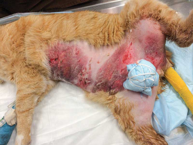
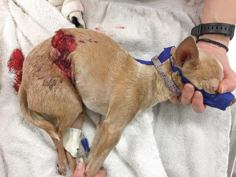
Wounds and fractures
Large wounds and degloving injuries can be overwhelming sights for the practitioner. It is important, however, to remember without significant associated bleeding, they represent very low immediate risk for the
patient’s life.
Wounds should be covered on presentation to prevent contamination and antibiotic treatment started promptly. As soon as the patient is stable, the wounds should be clipped, cleaned, and flushed. A new bandage should then be applied. Initial closure of the wound is rarely necessary and it is useful to reassess the wound the next day, allowing time for some tissues to declare themselves.
Fractures should be thoroughly evaluated on presentation, keeping in mind fractures can be a source of significant hemorrhage. An orthopedic exam should be performed on all limbs, checking for swelling, crepitation, instability, and pain. The spine should be evaluated for spinal fractures and a rectal exam should also be performed to discover pelvic fractures.
If possible, limb fractures distal to elbows and stifles should be stabilized using cast or splint. Radiographs should be delayed until the patient is stable enough to undergo deep sedation. If a spinal fractures if suspected, the animal should be kept immobilized on a rigid board.
into the abdominal cavity, and spinal fracture associated with paraplegia and loss of
deep pain sensation.
Prognosis
The animal trauma triage score
Prognostication has been improved with the use of various scoring systems. One is the animal trauma triage (ATT) score used to evaluate traumatic injuries. It is commonly used by emergency clinicians to assess the severity of a patient’s traumatic injuries and is designed to provide prognostic value based upon injury severity.
The ATT scoring system has six categories (perfusion, cardiac, respiratory, eye/muscle/integument, skeletal and neurologic) each of which is scored on a 0-3 scale (0 indicating slight to no injury and 3 indicating severe injury). Scores for each category are then added and the final score reflects injury severity. This scoring system is easy to use and relies only on initial physical exam and does not require any blood work or additional diagnostics.
In a study evaluating the ATT scoring system, survivors have been shown to have significantly lower ATT scores than non-survivors. The ATT score is also a significant predictor for likelihood of survival seven days after initial presentation.7
The ATT score represents a useful prognostic tool. However, it should never replace the initial triage and full physical assessment of the trauma patient although additional diagnostics are usually needed to fully assess and diagnose a trauma patient’s full injuries.
In addition to a good triage, the ABCD systematic approach helps the practitioner focus on potential life-threatening injuries and reduce the risk of overlooking internal injuries in polytrauma patients.
Virginie Wurlod, Dr. med vet, DACVECC, DECVECC, is an associate professor in small animal Emergency and Critical Care at Louisiana State University. Dr. Wurlod is originally from Switzerland, where she did her initial veterinary training, before pursuing an ECC residency in the U.S.
References
- Kolata, R. J., & Johnston, D. E. “Motor vehicle accidents in urban dogs: A study of 600 cases.” J Am Vet Med Assoc. 1975;167(10), pp. 938-941.
- Kolata, R. J., Kraut, N. H., & Johnston, D. E. “Patterns of trauma in urban dogs and cats: A study of 1,000 cases. J Am Vet Med Assoc. 1974; 164(5), pp. 499- 502.
- Simpson SA, Syring RS, Otto CM. “Severe blunt trauma in dogs: 235 cases (1997-2003).” J Vet Emerg Crit Care. (San Antonio). 2009; 19(6): pp. 588-602.
- Streeter EM, Rozanski EA, Laforcade-Buress A, et al. “Evaluation of vehicular trauma in dogs: 239 cases (January-December 2001).” J Am Vet Med Assoc. 2009; 235(4): pp. 405-408.
- Platt SR, Radaelli ST, McDonnell JJ. “The Prognostic Value of the Modified Glasgow Coma Scale in Head Trauma in Dogs.” J Vet Intern Med. 2001;15: pp. 581-584.
- Lisciandro GR, “Abdominal and thoracic focused assessment with sonography for trauma, triage, and monitoring in small anmals”. J Vet Emerg Crit Care. 2011;21(2): pp. 104-122.
- Rockar RA, Drobatz KS, “Development of a Scoring System For The Veterinary Trauma Patient.” J Vet Emerg Crit Care. 1994;2: pp. 77-83.
- Deborah C. Silverstein, Kate Hopper, Small Animal Critical Care Medicine. Elsevier, 2015, Chapter 1 Triage.
- Jamie M. Burkitt Creedon, Harold Davis, Advanced Monitoring and Procedures for Small Animal Emergency and Critical Care,” Wiley-Blackwell, 2012 Chapter 1 Triage.