In recent years, veterinary medicine has witnessed a remarkable transformation driven by advancements in equipment and a growing expectation to provide the best care possible for our patients.
There are three trends nationwide changing how general practices deliver care: minimally invasive surgery, ultrasound, and panoramic dental imaging. These equipment trends can help to deliver better patient care and potentially faster patient recovery. Additionally, utilization of these tools presents as a learning opportunity to veterinary technicians and allows general practice to offer more services.
Minimally invasive
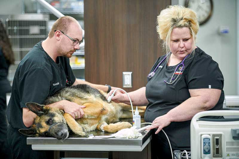
One of the most significant trends in veterinary medicine is the increasing use of minimally invasive surgery equipment. More veterinarians fresh out of school have been taught to use these tools and surgical procedures. From vessel sealers to scopes, minimally invasive equipment allows veterinarians to perform a wide range of procedures in-house, reducing the need for referrals to specialized facilities.
This trend spills over from human medicine, where laparoscopic surgeries and vessel sealers have become the standard. These procedures offer a more affordable care option and are fast becoming a staple in today’s practices.
Laparoscopic equipment, including endoscopes, allow the use of tiny ports rather than large incisions. Veterinarians can perform complex surgeries while minimizing patient recovery time. Some manufacturers have a laparoscopic bundle, making it easy to incorporate everything you need for laparoscopic surgery into your clinic.
Assembling all the laparoscopic equipment into one bundle saves practices space and the costs associated with purchasing each separately. A bundle can mean a practice can begin to see return on their investment quickly as well.
Les Meadowcroft, president of VetOvation, explains: “We provide a turnkey package so that they can enable their practice to perform minimally invasive surgery, whether it’s a laparoscopic spay, a gastropexy, a liver biopsy, a cryptorchid resection, video otoscopy–looking in an ear, going up a nose, going down the esophagus and into the stomach to be able to take a biopsy of tissue or pull out a foreign body. This equipment bundle also includes a vessel sealer.”
A vessel sealer can save surgeons a significant amount of time and is better for patient recovery. Joe Chow, DVM, of Rusty Ridge Animal Center has been able to cut pyometra surgery down to about 12 minutes; further, it’s less time for a patient to be under anesthesia.
According to Meadowcroft, the vessel sealer breaks down the hydrogen bonds in cells, making them go from a liquid state to a sticky coagulum.
“The vessel sealer enables veterinarians to clamp onto the tissue, then activate the energy that sends a signal to a generator,” Meadowcroft says. “Once that tissue is sealed, they’re able to cut with the blade. This enables them to do surgery faster, reduces blood loss for that patient and improves recovery time for that patient.”
Real-time assistance
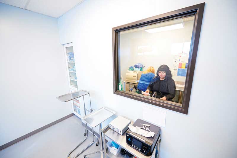
Ultrasound technology has long been a staple in veterinary diagnostics, but real-time assistance and training boomed during the pandemic, when clinics could not get on-site training. This innovation allows technicians to log into ultrasound systems remotely and assist with scanning procedures, making it convenient for the veterinary team.
In human medicine, trained technicians often handle ultrasound scans, and now the veterinary field is following suit. Technicians are trained to acquire images and upload them to be reviewed by veterinarians, freeing up valuable time for the doctors while ensuring the best in patient care.
While recent graduates may be familiar with ultrasound, there is an ever-expanding array of options, from handheld units practitioners can use with their phone, to advanced systems for cardiac use.
Students who are graduating from veterinary school are having more interaction with ultrasound during their school time. The equipment has moved away from being very difficult to use–a system with hundreds of buttons–to being touchscreen and operate very much like your phone.
With the ease of use of the systems and interactive training, ultrasound has gone from, “It’s going to take me six to eight months to be good at it,” to getting trained and in use quickly. With in-system training, practices can be up and running with ultrasound in no time.
“We go anywhere from wireless handheld ultrasounds for point of care, all the way to advanced ultrasounds for 4D cardiac,” shares Randy Laufersky, president of Core Imaging.
“We start out with our handheld ultrasound. It’s wireless. It will connect to any device, whether it be an Android or Apple. The great thing about this is we can use it for small animals, but if you need it to be used for a large animal, or you want to go from a micro convex to linear, you don’t need a whole new device. All you do is pop the head off, put on the new head and you’re ready to go. This is a great product because it has multiple uses and vet staff will not have to change the most expensive part.”
The next step up is a unit that is as easy to use as an iPad. A 13-in. screen gives staff access to high quality resolution, but also training. “Cloud access to this is incredible,” says Laufersky. “We can do customer service right through the system so that if something goes wrong, it’s not a week until you get service. It is literally minutes until we’re in the system and diagnosing issues.”
Another ultrasound is a cart-based, 19-in. touchscreen system with a seven-hour battery life. This unit is also as easy to use as a phone or tablet, and staff can go room to room without having to plug it in. Access to training is done through the unit as well.
When choosing an ultrasound, it is important to consider:
- User skill level
- Where you want to be with ultrasound in the next five years
- If education is something that’s a big portion of where you want to be
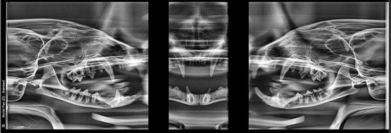
Panoramic X-ray system
Rather than taking an image tooth by tooth, which takes about 30 to 40 minutes, veterinarians and vet techs can get a comprehensive scan of an animal’s entire mouth in less than 20 seconds using a panoramic X-ray system.
This technology provides a visual aid for both veterinarians and pet owners, helping them have a better understanding of their pet’s dental health.
A built-in software allows for presets for different jaw sizes, from Chihuahuas to basset hounds. A laser light guides staff to position the patient correctly. Less time spent imaging means less time under anesthesia for the patient, and technicians can then proceed to doing the dental work after the scan.
The superior imaging is remarkable as well. During each yearly visit, a scan like this can visibly show, from year to year, progression of issues or disease. This happens on the human side, and we have come to expect it. Dental radiography is becoming more and more of a standard and expectation in veterinary medicine as well.
Saving time and gaining skills
Investing in new equipment helps veterinarians advance their practice, increase services offered for patients, and offer continued learning to staff.
These trends represent just a glimpse into the future of veterinary medicine. As technology continues to advance and veterinarians embrace these innovations, we can expect even more groundbreaking developments that will further enhance patient health and boost staff skills.
Shawn Gann is an equipment specialist with Patterson Veterinary and has been with the company for eleven years. He is based in the Tampa, Fla.